

A new mechanism of RhoA ubiquitination and degradation: roles of SCF(FBXL19) E3 ligase and Erk2
- PMID: 23871831
- PMCID: PMC3834026
- DOI: 10.1016/j.bbamcr.2013.07.005
A new mechanism of RhoA ubiquitination and degradation: roles of SCF(FBXL19) E3 ligase and Erk2
Abstract
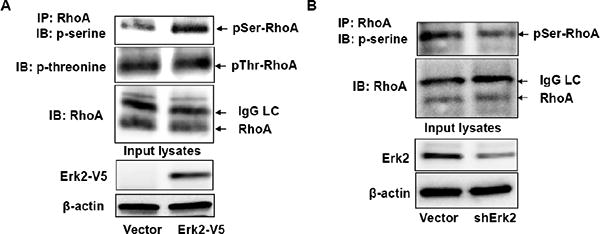
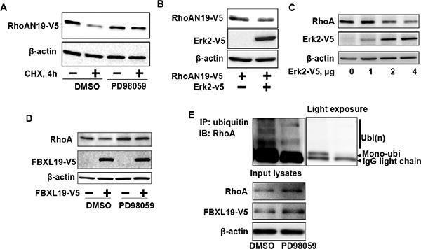
RhoA is a small GTPase multifunctional protein that regulates cell proliferation and cytoskeletal reorganization. Regulation of its protein stability plays an important role in its biological functions. We have shown that a Skp1-Cul1-F-box (SCF) FBXL19 E3 ubiquitin ligase targets Rac1, a related member of the Rho family for ubiquitination and degradation. Here, SCF(FBXL19) mediates RhoA ubiquitination and proteasomal degradation in lung epithelial cells. Ectopically expressed FBXL19 decreased RhoA wild type, active, and inactive forms. Cellular depletion of FBXL19 increased RhoA protein levels and extended its half-life. FBXL19 bound the small GTPase in the cytoplasm leading to RhoA ubiquitination at Lys(135). A RhoA(K135R) mutant protein was resistant to SCF(FBXL19)-mediated ubiquitination and degradation and exhibited a longer lifespan. Protein kinase Erk2-mediated phosphorylation of RhoA was both sufficient and required for SCF(FBXL19)-mediated RhoA ubiquitination and degradation. Thus, SCF(FBXL19) targets RhoA for its disposal, a process regulated by Erk2. Ectopically expressed FBXL19 reduced phosphorylation of p27 and cell proliferation, a process mediated by RhoA. Further, FBXL19 cellular expression diminished lysophosphatidic acid (LPA)-induced phosphorylation of myosin light chain (MLC) and stress fiber formation. Hence, SCF(FBXL19) functions as a RhoA antagonist during cell proliferation and cytoskeleton rearrangement. These results provide the first evidence of an F-box protein targeting RhoA thereby modulating its cellular lifespan that impacts cell proliferation and cytoskeleton rearrangement.
Keywords: Cell proliferation; Phosphorylation; Protein stability; Small GTPase protein; Stress fiber; Ubiquitin-proteasome system.
© 2013.
Figures


Similar articles
-
SCF E3 ligase F-box protein complex SCF(FBXL19) regulates cell migration by mediating Rac1 ubiquitination and degradation.FASEB J. 2013 Jul;27(7):2611-9. doi: 10.1096/fj.12-223099. Epub 2013 Mar 19. FASEB J. 2013. PMID: 23512198 Free PMC article.
-
SCF FBXW17 E3 ubiquitin ligase regulates FBXL19 stability and cell migration.J Cell Biochem. 2021 Apr;122(3-4):326-334. doi: 10.1002/jcb.29860. Epub 2020 Oct 14. J Cell Biochem. 2021. PMID: 33053230 Free PMC article.
-
Histone acetyltransferase CBP promotes function of SCF FBXL19 ubiquitin E3 ligase by acetylation and stabilization of its F-box protein subunit.FASEB J. 2018 Aug;32(8):4284-4292. doi: 10.1096/fj.201701069R. Epub 2018 Mar 9. FASEB J. 2018. PMID: 29522376 Free PMC article.
-
Role of SKP1-CUL1-F-box-protein (SCF) E3 ubiquitin ligases in skin cancer.J Genet Genomics. 2013 Mar 20;40(3):97-106. doi: 10.1016/j.jgg.2013.02.001. Epub 2013 Feb 10. J Genet Genomics. 2013. PMID: 23522382 Free PMC article. Review.
-
Ubiquitylation of active Rac1 by the E3 ubiquitin-ligase HACE1.Small GTPases. 2012 Apr-Jun;3(2):102-6. doi: 10.4161/sgtp.19221. Small GTPases. 2012. PMID: 22790197 Free PMC article. Review.
Cited by
-
Emerging roles of deubiquitinating enzymes in actin cytoskeleton and tumor metastasis.Cell Oncol (Dordr). 2024 Aug;47(4):1071-1089. doi: 10.1007/s13402-024-00923-z. Epub 2024 Feb 7. Cell Oncol (Dordr). 2024. PMID: 38324230 Review.
-
F-box proteins and cancer: an update from functional and regulatory mechanism to therapeutic clinical prospects.Theranostics. 2020 Mar 4;10(9):4150-4167. doi: 10.7150/thno.42735. eCollection 2020. Theranostics. 2020. PMID: 32226545 Free PMC article. Review.
-
miR-137-LAPTM4B regulates cytoskeleton organization and cancer metastasis via the RhoA-LIMK-Cofilin pathway in osteosarcoma.Oncogenesis. 2023 May 6;12(1):25. doi: 10.1038/s41389-023-00471-5. Oncogenesis. 2023. PMID: 37147294 Free PMC article.
-
The ubiquitin ligase RNF8 regulates Rho GTPases and promotes cytoskeletal changes and motility in triple-negative breast cancer cells.FEBS Lett. 2021 Jan;595(2):241-252. doi: 10.1002/1873-3468.13999. Epub 2020 Dec 5. FEBS Lett. 2021. PMID: 33205415 Free PMC article.
-
Regulation of Cdc42 protein turnover modulates the filamentous growth MAPK pathway.J Cell Biol. 2022 Dec 5;221(12):e202112100. doi: 10.1083/jcb.202112100. Epub 2022 Nov 9. J Cell Biol. 2022. PMID: 36350310 Free PMC article.
References
-
- Hall A. Rho GTPases and the actin cytoskeleton. Science (New York, NY. 1998;279:509–514. - PubMed
-
- Etienne-Manneville S, Hall A. Rho GTPases in cell biology. Nature. 2002;420:629–635. - PubMed
-
- Rossman KL, Der CJ, Sondek J. GEF means go: turning on RHO GTPases with guanine nucleotide-exchange factors. Nature reviews. 2005;6:167–180. - PubMed
-
- Kranenburg O, Poland M, Gebbink M, Oomen L, Moolenaar WH. Dissociation of LPA-induced cytoskeletal contraction from stress fiber formation by differential localization of RhoA. Journal of cell science. 1997;110(pt 19):2417–2427. - PubMed
-
- Sauzeau V, Le Mellionnec E, Bertoglio J, Scalbert E, Pacaud P, Loirand G. Human urotensin II-induced contraction and arterial smooth muscle cell proliferation are mediated by RhoA and Rho-kinase. Circulation research. 2001;88:1102–1104. - PubMed
Publication types
MeSH terms
Substances
Grants and funding
LinkOut - more resources
Full Text Sources
Other Literature Sources
Molecular Biology Databases
Research Materials
Miscellaneous
