Review
. 2013 Nov-Dec;1827(11-12):1407-27.
doi: 10.1016/j.bbabio.2013.07.006. Epub 2013 Jul 19.

Evolution of cytochrome bc complexes: from membrane-anchored dehydrogenases of ancient bacteria to triggers of apoptosis in vertebrates

Affiliations
Review

Evolution of cytochrome bc complexes: from membrane-anchored dehydrogenases of ancient bacteria to triggers of apoptosis in vertebrates

Daria V Dibrova et al. Biochim Biophys Acta. 2013 Nov-Dec.

Abstract

This review traces the evolution of the cytochrome bc complexes from their early spread among prokaryotic lineages and up to the mitochondrial cytochrome bc1 complex (complex III) and its role in apoptosis. The results of phylogenomic analysis suggest that the bacterial cytochrome b6f-type complexes with short cytochromes b were the ancient form that preceded in evolution the cytochrome bc1-type complexes with long cytochromes b. The common ancestor of the b6f-type and the bc1-type complexes probably resembled the b6f-type complexes found in Heliobacteriaceae and in some Planctomycetes. Lateral transfers of cytochrome bc operons could account for the several instances of acquisition of different types of bacterial cytochrome bc complexes by archaea. The gradual oxygenation of the atmosphere could be the key evolutionary factor that has driven further divergence and spread of the cytochrome bc complexes. On the one hand, oxygen could be used as a very efficient terminal electron acceptor. On the other hand, auto-oxidation of the components of the bc complex results in the generation of reactive oxygen species (ROS), which necessitated diverse adaptations of the b6f-type and bc1-type complexes, as well as other, functionally coupled proteins. A detailed scenario of the gradual involvement of the cardiolipin-containing mitochondrial cytochrome bc1 complex into the intrinsic apoptotic pathway is proposed, where the functioning of the complex as an apoptotic trigger is viewed as a way to accelerate the elimination of the cells with irreparably damaged, ROS-producing mitochondria. This article is part of a Special Issue entitled: Respiratory complex III and related bc complexes.

Keywords: Bioenergetics, molecular evolution, ubiquinol:cytochrome c oxidoreductase; cardiolipin, cell death, photosynthesis, apoptosome; cytochrome c; plastoquinone; ubiquinone.


Figures

Figure 1
Figure 1
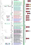
Phylogenetic tree of cytochromes b. Each protein is indicated by its NCBI’s GenInfo identifier (gi number), followed by the name of the source organism; in two instances, the proteins are labeled by their PDB codes (1VF5 and 1NTM). The colors of protein names indicate the taxonomical positions of the respective species. The detailed correspondence between colors and taxons is provided in Table S1 of Supplementary Materials (File 1). Black dots mark branches with strong bootstrap support > 70%, white dots show branches with good bootstrap support > 50%. Alternative schematic representations of the same tree are given as Fig. S2 in File 1 of Supplementary Materials (with indicated chemical nature of the predominant pool quinone) and also provided as a separate File 2 of Supplementary Materials (with bootstrap values and aLRT results indicated). Clades marked on the trees are as follows: A, cyanobacterial and plant clade; eukaryotic sequences are marked by thick orange branches; B, clade with cytochrome b of Heliobacterium modesticaldum and related proteins; C, clade of Bacilli members and Thermodesulfovibrio yellowstonii; D and G, unusual clades, see the main text; E, mostly archaeal clade; F, clade with sequences from Deinococcus-Thermus bacteria, actinobacteria and haloarchaea; H, fusions between cytochrome b and different sets of redox domains; I, proteobacterial clade with mitochondrial cytochromes b and proteins from Aquificae (the mitochondrial cytochrome is indicated by an orange-colored branch), J, Chlorobi clade. Specific marks are as follows: 1) Complexes with subunit IV as a separate protein are marked with symbol -/ /-. The gi’s of the pairs “cytochrome b - subunit IV” are separated with a space (for instance, “7525063 64” means that cytochrome b6-like and subunit IV-like proteins have gi’s 7525063 and 7525064, respectively). 2) In the heme cn binding motif, Cys residue is shown in orange, Gly residues are shown in green and other residues are marked grey. 3) Squares with different filling show the following deviations from the typical quinone-binding motif P[DE]W[FY] in the subunit IV (or in the C-terminal part of the “long” cytochrome b): black square, PVW[FY], green square, PPW[FY], gray square, PDIY, white square, P W[FY], white square with a cross, G WF, red square with a cross, [LV]DW[FY], red square with a slash, FDW[FY]. 4) The red cross symbol indicates the absence of subunit IV. 5) If the genome contains sequences coding for unusual cytochromes ñ (see the main text), the proteins from this genome are marked with the “red plus” sign. 6) Lines of different colors before the names of the proteins indicate different conservative linkers between the cytochrome b6-like parts and subunit IV-like parts of the full-length cytochromes b. Frames of the same color to the left show sequence logo diagrams for these linkers. The sequences 118575215 (Cenarchaeum symbiosum) and 161529051 (Nitrosopumilus maritimus) lack linker parts. 7) Figures after the species names depict divergence from the typical structure shown in a rectangle in the top right corner. Four red rectangles correspond to a 4-helical bundle (cytochrome b6-like part), two dark red rectangles depict two well-aligned helices of the subunit IV, hatched dark rectangles indicate an unaligned third helix of subunit IV. Vertical grey rectangles correspond to additional helices after subunit IV, yellow rectangles with round edges signify domains with heme-binding sites (cytochrome c-like domains), the small grey rectangle with round edges indicates a small domain conserved in actinobacteria. Finally, rectangles with marks “F1” and “F2” in clade H indicate two types of fusions with different sets of domains (see File 1 of Supplementary materials).
Figure 2
Figure 2
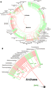
Presence and absence of cytochrome b (COG1290), used as a marker of the cytochrome bc1 complexes, mapped on the ribosomal protein-based phylogenetic tree of prokaryotes [233, 234]. Branch lengths do not exactly reflect the evolutionary distance between the nodes. The assignment of proteins from the RefSeq release 45 (Jan 07, 2011) to the Clusters of Orthologous Groups (COGs) [236] was taken from the NCBI FTP site (ftp://ftp.ncbi.nih.gov/pub/wolf/COGs/Prok1202/). The redundancy in the list of complete genomes from the RefSeq release 45 was reduced by manually removing species of the same genera, which resulted in a list of 582 prokaryotic species. Taxonomy data from the NCBI (http://www.ncbi.nlm.nih.gov/taxonomy) [235] to the level of family were used to map the taxonomy for these genomes on the aforementioned large-scale tree. For calculations, the set of 582 bacterial genomes was further reduced to a compact set of 102 bacterial genomes. Within bacteria, we selected genomes which contained cytochromes b (68 genomes), as well as genomes of closely related species which did not contain cytochrome b. In calculations with the COUNT software, a full sample of 115 archaeal genomes and a compact set of 102 bacterial genomes from all major phyla were used, which resulted in a sample of 217 genomes. 29 COGs which occur in at least half of major bacterial and archaeal phyla and do not contain more than 10 members in each genome were selected randomly. These COGs were used as a reference for the estimation of typical rates of gene losses, gene gains and other parameters in COUNT by the “Gain-Loss-Duplication” model with default parameters. For the reference COGs and the cytochrome b COG1290, respectively, the occurrences in each of 217 sampled genomes were calculated as described above. The rates of gene losses and gains were optimized on a subset of 217 genomes chosen to satisfy the computational requirements of the program. (A) Phylogenetic tree of prokaryotes. For the phyla that contain cytochromes b, the letters in brackets indicate the clades in Figure 1 that include cytochrome b sequences found in these phyla. (B) Enlarged archaeal clade. The estimated probabilities of independent acquisition of cytochrome(s) b in each group are given in square brackets.
Figure 3
Figure 3
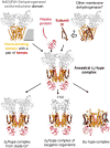
Evolutionary scenario for the cytochrome bc complexes. Cytochrome b6-like parts (the 4-helical bundle) are colored orange, subunit IV-like parts are colored dark red, the Rieske proteins are colored pink. The three-helix subunit IV is arbitrarily suggested to be recruited from a membrane dehydrogenase, see text for further details.
Figure 4
Figure 4
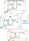
Q-cycle mechanisms in different cytochrome bc complexes. A, a menaquinone (MQ)-dependent b6f-type complex of anoxic organisms; B, a plastoquinone (PQ)-dependent b6f-type of oxygenic organisms; C, an ubiquinone (UQ)-dependent bc1–type complex of aerobic organisms; see text for further details and references.
Figure 5
Figure 5
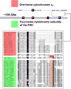
Comparison of the one-heme cytochome c2 from Rhodopseudomonas palustris and its closest homologs (colored red) and the four-heme PRC cytochome subunit from Blastochloris viridis and its homologs (colored green). The heme-binding sites are marked by red rectangles. The part of the multiple alignment between C-terminal parts of the proteins is shown below in the box. The black stretches are the regions which are not aligned on multiple alignment, while the blue stretches show regions with detectable similarity that are included in the multiple alignment.
Figure 6
Figure 6
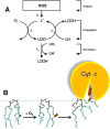
Oxidation of cardiolipin in the mitochondrial membrane. A, general scheme of lipid peroxidation, adapted from [237]; L is a lipid molecule, QH2and QH are membrane ubiquinol and its semiquinone form, respectively. B, transformations of a cardiolipin molecule upon peroxidation according to [177, 178]. The black dot indicates the position of an unpaired electron, see the main text for further details.
Figure 7
Figure 7
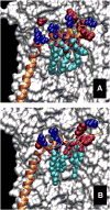
Clusters of occluded cardiolipin molecules in the cytochrome bc1 complexes. Cardiolipin molecules are colored by element: carbon – cyan, oxygen – red and phosphorus – yellow. The 9.5 kD subunit (subunit G in the bovine bc1 and subunit H in the yeast bc1) is colored orange, its positively charged residues are colored blue. The positively charged residues, provided by cytochromes b and c1, are colored violet, see also Figs S7–S11 in Supplementary Materials. A, bovine cytochrome bc1 complex, PDB record 1PP9 [202]; B, yeast cytochrome bc1 complex, PDB record 3CX5 [189]. The figure was produced with the help of the VMD software package [238].
Figure 8
Figure 8
Triggering of the intrinsic apoptotic pathway by a mitochondrion in a mammalian cell. The ROS occasionally generated in the bc1 (red dashed arrow), by oxidizing cardiolipin and eventually damaging the bc1 itself, produce two potent sources of ROS, thus accelerating the triggering of the apoptosis (see the main text for further details). Potential positive feedback loops, namely the increase in the yield of ROS upon damaging of the bc1 [159] and the transformation of cytochrome c into a peroxidase by oxidized cardiolipin (CL) molecules [177, 178] are emphasized. See the text for further details.

Similar articles

Cited by

References

    1. Berry EA, Guergova-Kuras M, Huang LS, Crofts AR. Structure and function of cytochrome bc complexes. Ann Rev Biochem. 2000;69:1005–1075. - PubMed
    1. Cramer WA, Hasan SS, Yamashita E. The Q cycle of cytochrome bc complexes: a structure perspective. Biochim Biophys Acta. 2011;1807:788–802. - PMC - PubMed
    1. Al-Attar S, de Vries S. Energy transduction by respiratory metallo-enzymes: From molecular mechanism to cell physiology. Coord Chem Rev. 2012;257:64–80.
    1. Berry EA, De Bari H, Huang LS. Unanswered questions about the structure of cytochrome bc complexes. Biochim Biophys Acta. 2013 doi: 10.1016/j.bbabio.2013.04.006. - DOI - PubMed
    1. Hasan SS, Yamashita E, Cramer WA. Transmembrane signaling and assembly of the cytochrome bf-lipidic charge transfer complex. Biochim Biophys Acta. 2013 doi: 10.1016/j.bbabio.2013.03.002. - DOI - PMC - PubMed

Publication types

MeSH terms

LinkOut - more resources