Review
. 2013 Aug;34(3):211-27.
doi: 10.1016/j.yfrne.2013.07.003. Epub 2013 Jul 17.

Modulation of body temperature and LH secretion by hypothalamic KNDy (kisspeptin, neurokinin B and dynorphin) neurons: a novel hypothesis on the mechanism of hot flushes

Affiliations
Review

Modulation of body temperature and LH secretion by hypothalamic KNDy (kisspeptin, neurokinin B and dynorphin) neurons: a novel hypothesis on the mechanism of hot flushes

Naomi E Rance et al. Front Neuroendocrinol. 2013 Aug.

Abstract

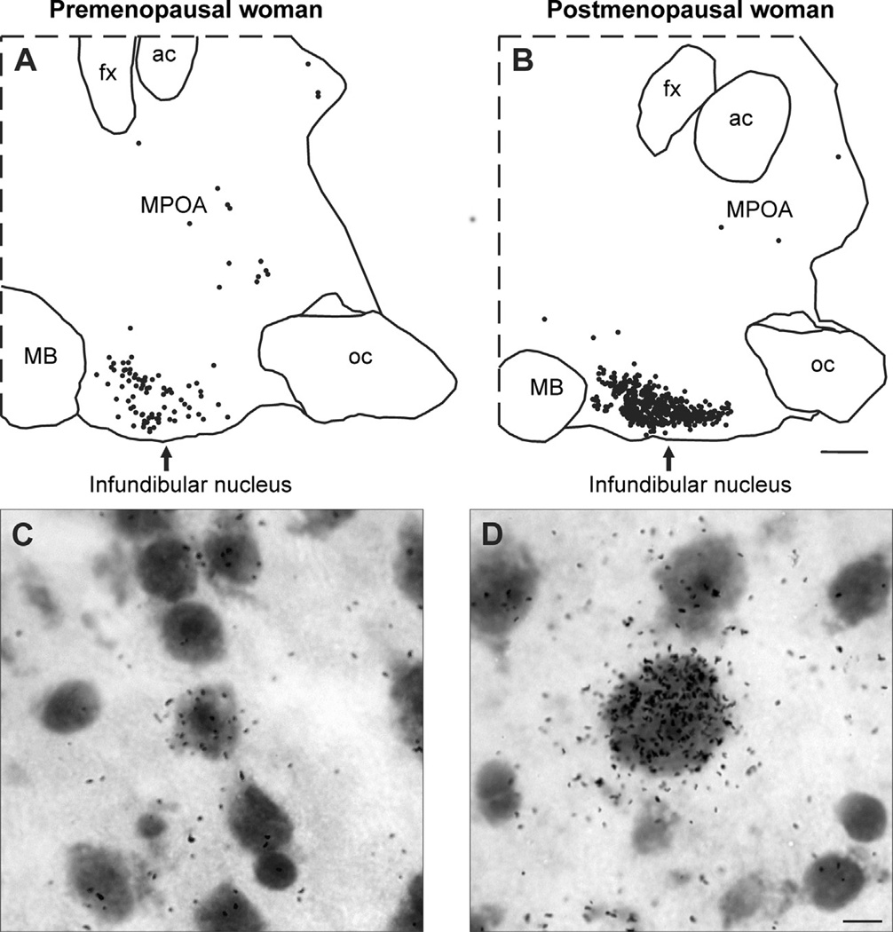
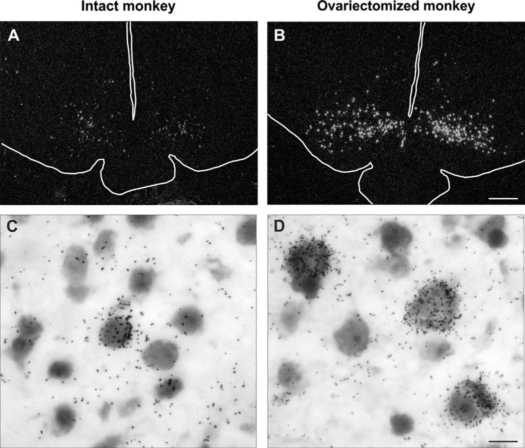
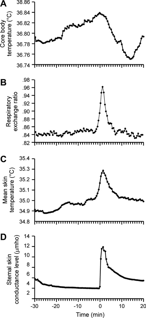
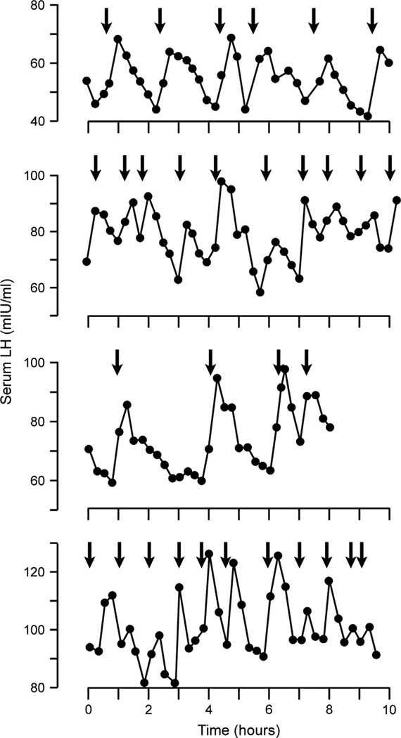
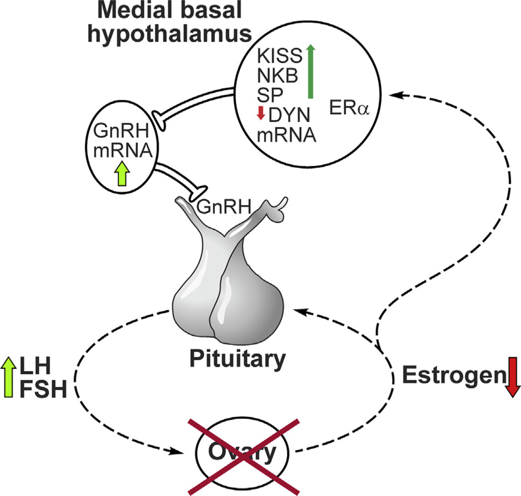
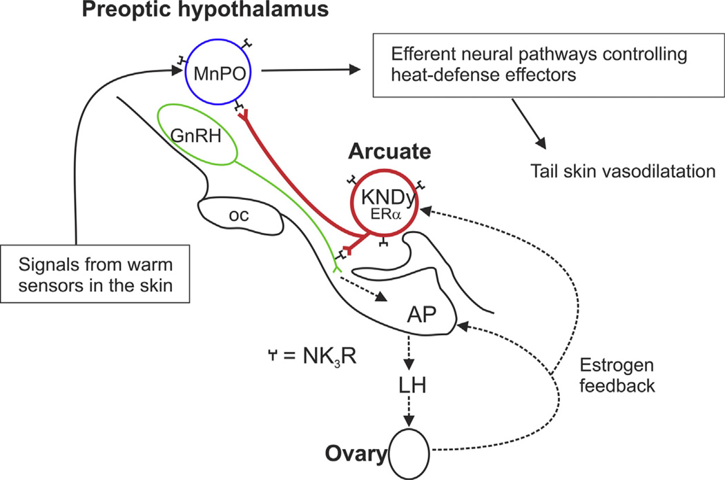
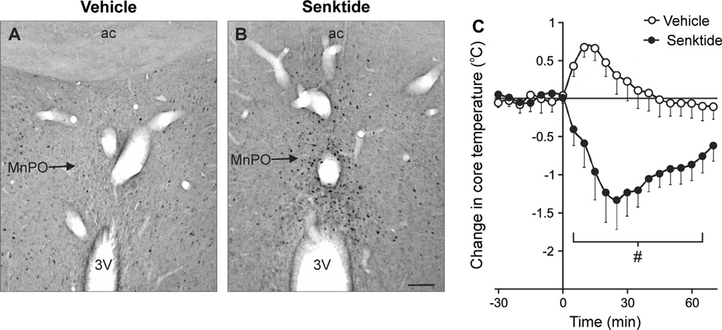
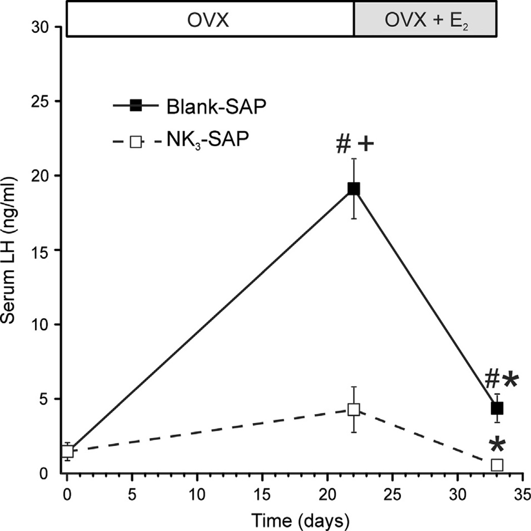
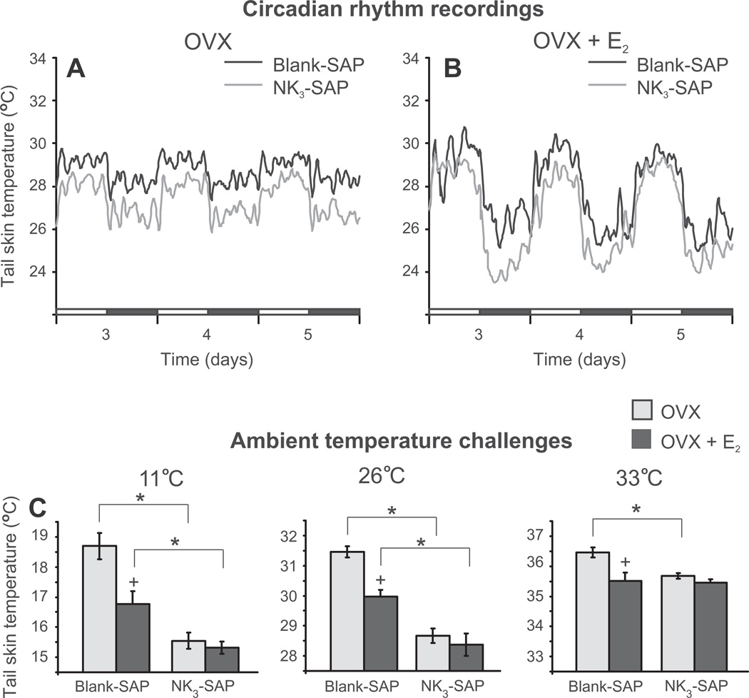
Despite affecting millions of individuals, the etiology of hot flushes remains unknown. Here we review the physiology of hot flushes, CNS pathways regulating heat-dissipation effectors, and effects of estrogen on thermoregulation in animal models. Based on the marked changes in hypothalamic kisspeptin, neurokinin B and dynorphin (KNDy) neurons in postmenopausal women, we hypothesize that KNDy neurons play a role in the mechanism of flushes. In the rat, KNDy neurons project to preoptic thermoregulatory areas that express the neurokinin 3 receptor (NK3R), the primary receptor for NKB. Furthermore, activation of NK₃R in the median preoptic nucleus, part of the heat-defense pathway, reduces body temperature. Finally, ablation of KNDy neurons reduces cutaneous vasodilatation and partially blocks the effects of estrogen on thermoregulation. These data suggest that arcuate KNDy neurons relay estrogen signals to preoptic structures regulating heat-dissipation effectors, supporting the hypothesis that KNDy neurons participate in the generation of flushes.

Keywords: Estrogen; GnRH; LH; Menopause; Reproduction; Thermoregulation.


Figures

Fig. 1
Fig. 1
(A and B) Computer-assisted maps of kisspeptin mRNA-expressing neurons in sagittal hypothalamic sections from a premenopausal (A) and a postmenopausal (B) woman hybridized with cDNA probes complimentary to kisspeptin mRNA and visualized using autoradiography. Each symbol represents one labeled neuron. In the infundibular nucleus of postmenopausal women, there is a marked increase in the number of neurons expressing kisspeptin mRNA. Small numbers of kisspeptin neurons are also identified within the MPOA, but these did not change in postmenopausal women. (C and D) Photomicrographs of kisspeptin mRNA-expressing neurons in the infundibular nucleus of a premenopausal (C) or postmenopausal (D) woman. In postmenopausal women, kisspeptin neurons are hypertrophied and display increased numbers of audioradiographic grains, indicative of increased gene expression. Nearly identical changes can be seen in NKB neurons in the infundibular nucleus of postmenopausal women (Rance and Young, 1991). Scale Bar = 2 mm in B and applies to A and B. Scale bar = 10 µm in D and applies to C and D. ac, anterior commissure; fx, fornix; MB, mammillary body; MPOA, medial preoptic area; oc, optic chiasm Modified with permission from Rometo et al. (2007).
Fig. 2
Fig. 2
(A and B) Darkfield photomicrogaphs of coronal hypothalamic sections from an intact (A) or ovariectomized (B) cynomolgus monkey hybridized with cDNA probes complimentary to NKB mRNA (Tac3 in the monkey) and visualized using autoradiography. The outlines of the base of the brain, pituitary stalk and third ventricle have been drawn on these photomicrographs. Note the marked increase in the number of labeled neurons in arcuate nucleus of the ovariectomized monkey. (C and D) Brightfield photomicrogaphs of neurons expressing NKB mRNA visualized using in situ hybridization in the arcuate nucleus of an intact (C) or ovariectomized (D) cynomolgus monkey. The increase in cell size in the ovariectomized monkeys is accompanied by increased numbers of labeled cells and autoradiographic grains/neuron, indicative of a marked increase in gene expression in response to ovariectomy. These findings mimic the changes in NKB cell size and gene expression observed in the infundibular nucleus of postmenopausal women. Scale bar = 0.5 mm in B (applies to A and B), Scale bar = 25 µm in D (applies to C and D). Modified with permission from Sandoval-Guzmán et al. (2004).
Fig. 3
Fig. 3
(A) Average core body temperature, (B) respiratory exchange ratio (C) skin temperature and (D) sternal skin conductance recorded from 29 hot flushes in 14 postmenopausal women. Time 0 is the onset of the rise in sternal skin conductance. Vasodilatation and sweating (reflected in C and D, respectively) are physiological heat-dissipation effectors that are activated during hot flushes. A small increase in core temperature is seen before the flush, followed by a larger drop due to the activation of heat-dissipation effectors (A). Adapted with permission from Freedman (1998).
Fig. 4
Fig. 4
Pattern of pulsatile LH release and associated flush episodes in four postmenopausal women. The arrows indicate flush onset. Each graph illustrates a separate 8- to 10-h study in which blood samples were obtained at 15-min intervals. The close timing of LH pulses and hot flushes provides evidence that the mechanism of flushes is linked with the hypothalamic control of pulsatile GnRH secretion. Reproduced with permission from Casper et al. (1979).
Fig. 5
Fig. 5
Schematic diagram showing the relationship between KNDy neurons and the neuroendocrine circuits controlling LH secretion in postmenopausal women. Degeneration of ovarian follicles results in loss of estrogen secretion (Hansen et al., 2008). Removal of estrogen negative feedback increases LH secretion from the anterior pituitary gland into the systemic circulation. In the infundibular nucleus, there is hypertrophy of a subpopulation of neurons expressing ERα, kisspeptin, NKB and substance P mRNA accompanied by increased kisspeptin, NKB and substance P gene expression (Rance and Young, 1991; Rometo et al., 2007; Rometo and Rance, 2008; Sandoval-Guzmán et al., 2004). Neurons expressing dynorphin mRNA also exhibit neuronal hypertrophy, but the number of neurons expressing dynorphin gene transcripts is reduced (Rometo and Rance, 2008). Dual-labeled NKB/kisspeptin fibers are closely apposed to GnRH cell bodies and processes in the human medial basal hypothalamus (Hrabovszky et al., 2011). GnRH mRNA is increased in a separate subpopulation of neurons in the medial basal hypothalamus of postmenopausal women (Rance and Uswandi, 1996). Reproduced with permission from Rance (2009).
Fig. 6
Fig. 6
Typical records of core temperature (Tc, dotted line), tail skin temperature (Tsk, solid line) and ambient temperature (Ta dashed line) from three different Zucker lean rats at ambient temperatures of 27 °C (left), 29.5 °C (middle), or 32 °C (right). Note the large-amplitude fluctuations in tail skin temperature that occur at 29.5 °C, characteristic of tail skin vasomotion within the thermoneutral zone. Reproduced with permission from Romanovsky et al. (2002).
Fig. 7
Fig. 7
Effects of subcutaneous capsules of 17β-estradiol (E2) on average (±SEM) tail skin temperature of ovariectomized rats during the dark (A) or light (B) phase (n = 8–10 rats/group). In the dark phase (A), E2 treatment of ovariectomized (OVX) rats markedly reduces tail skin temperature from experimental day 2 through the end of the experiment. The reduction in tail skin temperature by E2 in the light phase (B) is delayed and smaller in magnitude than that seen in the dark phase. The acute drop in tail skin temperature during the light phase of days 0 and 2 (arrows) reflects sympathetic vasoconstriction at the time of capsule implantation. (C) Recording of tail skin temperature of an individual intact rat during the estrous cycle (moving average of 5 data points). This recording illustrates circadian rhythms, large fluctuations during most of the cycle and decreased tail skin temperature with lower fluctuations on proestrous night. The black bars represent the dark phase. * Significantly different in OVX + E2 rats compared to OVX. Reproduced with permission from Williams et al. (2010).
Fig. 8
Fig. 8
(A) Average (±SEM) core temperature of ovariectomized (OVX) and OVX + estradiol−17β (OVX + E2) rats at ambient temperatures from 13 °C to 34 °C (n = 11–12 rats/group). At most ambient temperatures, estradiol has no effect on core temperature. However, at ambient temperatures above 32.5 °C, the core temperature of OVX rats is significantly higher than OVX + E2 rats. These data show that estradiol-17β treatment of OVX rats improves core temperature regulation during heat exposure. (B) Heat loss index (HLI) range of OVX and OVX + E2 rats at ambient temperatures from 13 °C to 34 °C. HLI is a measure of tail skin vasomotion.An increase in HLI range indicates increased fluctuations in tail skin vasomotion, a characteristic finding within the thermoneutral zone (see Fig. 6 and Romanovsky et al., 2002). The straight line and parabola were fitted to these points using breakpoint analysis. The range of the parabola approximates the thermoneutral zone. These data show a shift in thermoneutral zone to lower ambient temperatures in OVX rats compared to OVX + E2 rats. The circles represent median values of 11–12 rats/group. * Significantly different OVX vs OVX + E2#Non-overlapping 95% confidence intervals comparing the first breakpoint for OVX vs. OVX + E2 rats. Adapted with permission from Dacks and Rance (2010).
Fig. 9
Fig. 9
Schematic diagram showing the relationship between KNDy neurons, GnRH neurons and the heat-defense pathway in the rat. KNDy neurons branch and project to GnRH terminals in the median eminence and preoptic structures that regulate body temperature (Krajewski et al., 2005, 2010; Nakamura and Morrison, 2010; Romanovsky et al., 2009; Tanaka et al., 2009; Yeo and Herbison, 2011; Yoshida et al., 2009). Secretion of GnRH into portal capillaries stimulates LH secretion from the anterior pituitary gland, which stimulates the secretion of estrogen (E2) from the ovaries. E2 negative feedback reduces serum LH and decreases NKB and kisspeptin mRNA in KNDy neurons (Rance and Bruce, 1994; Smith et al., 2005a). ERa, the isoform required for estrogen negative feedback (Dorling et al., 2003), is expressed in arcuate KNDy neurons (Burke et al., 2006) but not GnRH neurons (Hrabovszky et al., 2001). NK3R is expressed on arcuate KNDy neurons (Burke et al., 2006) and GnRH terminals in the median eminence (Krajewski et al., 2005). GnRH neurons express kisspeptin receptor mRNA (Han et al., 2005), but the location of the kisspeptin receptor protein on GnRH neurons has not been described. MnPO neurons express NK3R. The MnPO receives information from warm-sensitive, cutaneous thermoreceptors and project to CNS structures to modulate heat-dissipation effectors (Nakamura and Morrison, 2010; Yoshida et al., 2009). Not shown are KNDy neuron projections to the medial preoptic area, which contains warm-sensitive GABAergic neurons which may also express NK3R (Eberwine and Bartfai, 2011). Modified with permission from (Mittelman-Smith et al., 2012a).
Fig. 10
Fig. 10
(A and B) Photomicrographs of Fos expression in the MnPO, 90 min after focal microinfusion of either vehicle (A, aCSF) or senktide (B). Note the marked increase in Fos-immunoreactive nuclei in senktide-microinfused rats. (C) Core temperature before and after vehicle or senktide microinfusion into the MnPO of ovariectomized, estradiol-treated rats (ambient temperature of 21.5 °C, mean ± SEM, n = 6–8 microinfusions/group). Senktide microinfusion into the MnPO markedly decreased core temperature. Scale bar in B = 100 microns and applies to A and B. aCSF, artificial cerebrospinal fluid, 3V, Third ventricle; ac, anterior commissure; MnPO, median preoptic nucleus. # Significantly different, senktide compared with vehicle. Modified with permission from Dacks et al. (2011a).
Fig. 11
Fig. 11
Effects of KNDy neuron ablation on Serum LH (mean ± SEM, n = 6–8 rats/ group). On day 0, rats were ovariectomized (OVX) and injected with NK3-SAP (KNDy ablated) or Blank-SAP in the arcuate nucleus. From 20 to 23 days later and they were implanted s.c. with E2 capsules and sacrificed 11 days later. In Blank-SAP controls, serum LH was markedly increased after ovariectomy and reduced by E2 treatment. In KNDy-ablated rats, LH did not significantly rise three weeks after OVX. Serum LH was also lower in KNDy ablated rats, vs Blank-SAP controls after estrogen treatment. These data show that KNDy neurons are essential for the rise in LH secretion after ovariectomy and tonic LH secretion in estrogen-treated rats. # Significantly different, Blank-SAP vs. NK3-SAP. + Significantly different from intact values on day 0. * Significantly reduced after E2 treatment (compared to OVX). Modified with permission from Mittelman-Smith et al. (2012b).
Fig. 12
Fig. 12
(A and B) Circadian rhythms of tail skin temperature in KNDy-ablated (NK3-SAP) and control (Blank-SAP) rats that are ovariectomized (OVX, A) or OVX and treated with 17β-estradiol (OVX + E2, B). Circadian rhythms of tail skin temperature and activity were unchanged, but tail skin temperature was consistently lower in KNDy ablated rats than Blank-SAP controls. In both KNDy-ablated and control rats, estradiol decreased tail skin temperature during the dark phase (compare A and B). (C) Average tail skin temperature (±SEM) of KNDy-ablated (NK3-SAP) and control (Blank-SAP) rats subjected to different ambient temperatures in an environmental chamber (during the light phase). At the ambient temperature of 11 °C and 26 °C, the tail skin temperature of NK3-SAP animals was significantly lower than in Blank-SAP animals. Tail skin temperature of the Blank-SAP rats was reduced by E2 at all ambient temperatures, but not lowered by E2 treatment in NK3-SAP rats. The lower levels of tail skin temperature in KNDy-ablated rats provides evidence that KNDy neurons facilitate cutaneous vasodilatation, a cardinal sign of a hot flush. * Significantly different, NK3-SAP vs Blank-SAP. + Significantly different, OVX vs OVX+ 17β-estradiol. Adapted with permission from Mittelman-Smith et al. (2012a).

References

    1. Abel TW, Rance NE. Stereologic study of the hypothalamic infundibular nucleus in young and older women. J. Comp. Neurol. 2000;424:679–688. - PubMed
    1. Abel TW, Voytko ML, Rance NE. The effects of hormone replacement therapy on hypothalamic neuropeptide gene expression in a primate model of menopause. J. Clin. Endocrinol. Metab. 1999;84:2111–2118. - PubMed
    1. Alçin E, Sahu A, Ramaswamy S, Hutz ED, Keen KL, Terasawa E, Bethea CL, Plant TM. Ovarian regulation of kisspeptin neurones in the arcuate nucleus of the rhesus monkey (Macaca mulatta) J. Neuroendocrinol. 2013;25:488–496. - PMC - PubMed
    1. Alfinito PD, Huselton C, Chen X, Deecher DC. Pharmacokinetic and pharmacodynamic profiles of the novel serotonin and norepinephrine reuptake inhibitor desvenlafaxine succinate in ovariectomized Sprague-Dawley rats. Brain Res. 2006;1098:71–78. - PubMed
    1. Almeida MC, Steiner AA, Branco LG, Romanovsky AA. Cold-seeking behavior as a thermoregulatory strategy in systemic inflammation. Eur. J. Neurosci. 2006a;23:3359–3367. - PubMed

Publication types

MeSH terms