

Comment
doi: 10.1093/brain/awt154.
Epub 2013 Jul 19.
Microcysts in the inner nuclear layer from optic atrophy are caused by retrograde trans-synaptic degeneration combined with vitreous traction on the retinal surface
Affiliations
- PMID: 23872368
- PMCID: PMC3808684
- DOI: 10.1093/brain/awt154
Item in Clipboard
Comment
Microcysts in the inner nuclear layer from optic atrophy are caused by retrograde trans-synaptic degeneration combined with vitreous traction on the retinal surface
Brain.
2013 Nov.
No abstract available
Figures
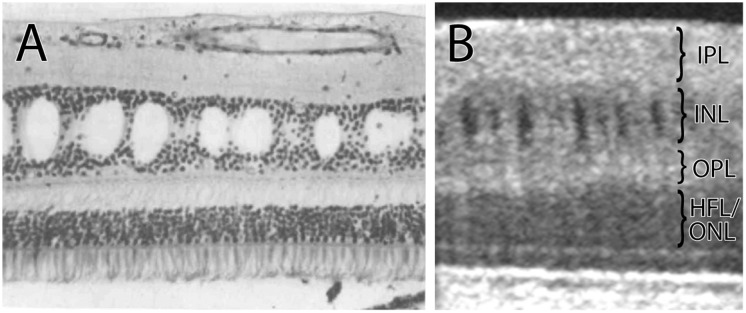
(A) Histological section of the retina showing cysts in the inner nuclear layer following retrograde trans-synaptic degeneration from a tumour of the optic nerve. Reproduced from Gills and Wadsworth (1967) with permission from James P. Gills. (B) Image obtained by optical coherence tomography from a patient with optic atrophy, showing cysts in the inner nuclear layer. IPL = inner plexiform layer; INL = inner nuclear layer; OPL = outer plexiform layer; HFL = Henle fibre layer; ONL = outer nuclear layer.
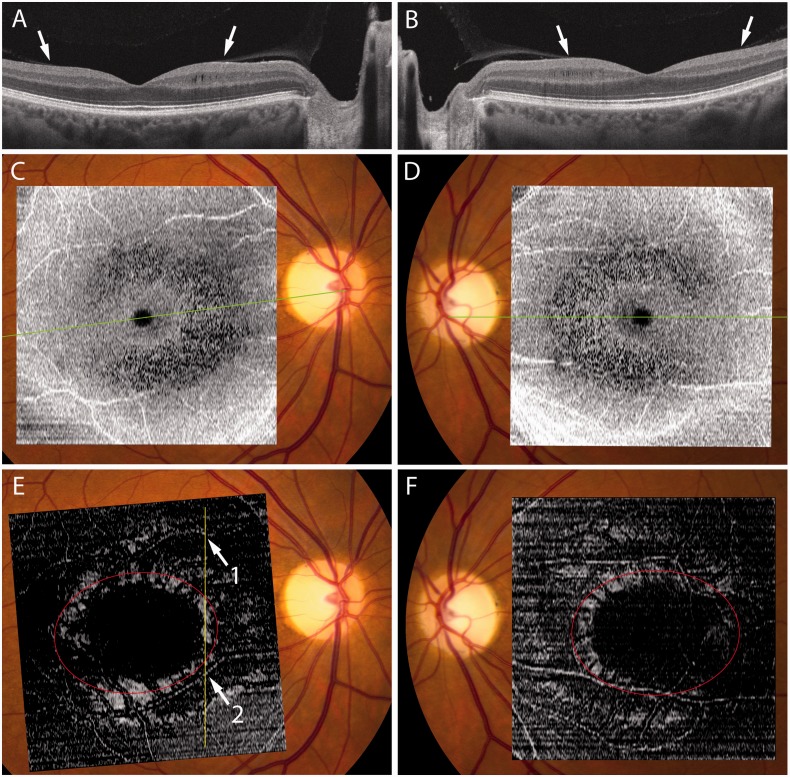
(A and B) Optical coherence tomography scans through the optic nerve and fovea in each eye of a 49-year-old female with dominant optic atrophy showing vitreous insertion (arrows). Nasally, but not temporally, vitreous traction has thickened the inner nuclear layer and produced microcysts. Note severe attenuation of the ganglion cell and nerve fibre layer. (C and D) Ring of microcysts between 3–8° visualized in en face reconstructions of 512 × 128 macular cubes achieved using Cirrus HD-OCT advanced visualization software (version 6.0) with a 68 µm RPE-based contour passing through the inner nuclear layer. The green lines correspond to the orientation of the scans shown above. (E and F) 20 µm internal limiting membrane based slab showing a ring of hyper-reflectivity where the retinal surface is perpendicular to incident light, presumably from vitreous traction. Red oval marks the insertion of the posterior hyaloid membrane, determined from inspection of serial cross-sections. In E, the vertical yellow line corresponds to the orientation of Fig. 3, with two prominent blood vessels denoted as 1 and 2.
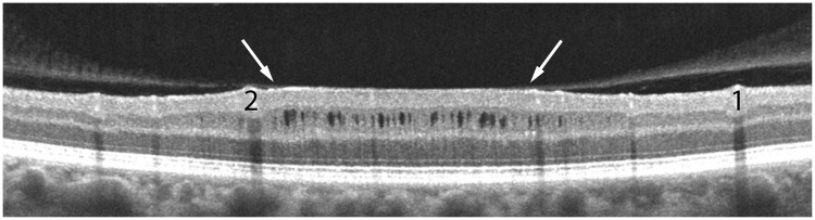
Optical tomography coherence scan of the right eye, corresponding to the yellow line in Fig. 2E with blood vessels marked 1 and 2, showing tenting of the inner nuclear layer from vitreous traction (arrows). The thickness of the inner nuclear layer is increased by 40% in the zone where it is elevated by traction, giving rise to ∼30 microcysts.
Comment in
-
Reply: Microcysts in the inner nuclear layer from optic atrophy are caused by retrograde trans-synaptic degeneration combined with vitreous traction on the retinal surface.Brain. 2013 Nov;136(Pt 11):e261. doi: 10.1093/brain/awt155. Epub 2013 Jul 19. Brain. 2013. PMID: 23872367 No abstract available.
Comment on
-
Microcystic macular oedema in multiple sclerosis is associated with disease severity.Brain. 2012 Jun;135(Pt 6):1786-93. doi: 10.1093/brain/aws098. Epub 2012 Apr 25. Brain. 2012. PMID: 22539259 Free PMC article.
References
-
- Abegg M, Zinkernagel M, Wolf S. Microcystic macular degeneration from optic neuropathy. Brain. 2012;135:e225. - PubMed
-
- Barboni P, Carelli V, Savini G, Carbonelli M, La Morgia C, Sadun AA. Microcystic macular degeneration from optic neuropathy: not inflammatory, not trans-synaptic degeneration. Brain. 2013 Advance Access published on February 8, 2013. - PubMed
-
- Gills JP, Wadsworth JA. Retrograde transsynaptic degeneration of the inner nuclear layer of the retina. Invest Ophthalmol Vis Sci. 1967;6:437–48.
-
- Jindahra P, Petrie A, Plant GT. Retrograde trans-synaptic retinal ganglion cell loss identified by optical coherence tomography. Brain. 2009;132:628–34. - PubMed
Publication types
MeSH terms
Grants and funding
LinkOut - more resources
Full Text Sources
Other Literature Sources
Medical
