. 2013 Oct;135(2):414-24.
doi: 10.1093/toxsci/kft154. Epub 2013 Jul 19.

Macrophage solubilization and cytotoxicity of indium-containing particles in vitro

Affiliations

Macrophage solubilization and cytotoxicity of indium-containing particles in vitro

William M Gwinn et al. Toxicol Sci. 2013 Oct.

Abstract

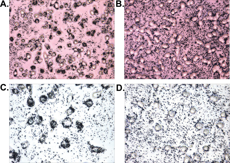
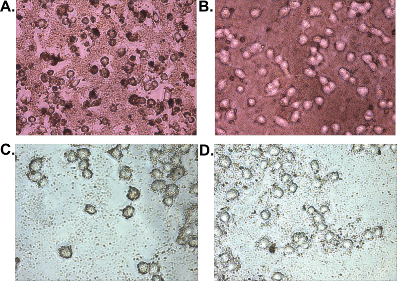
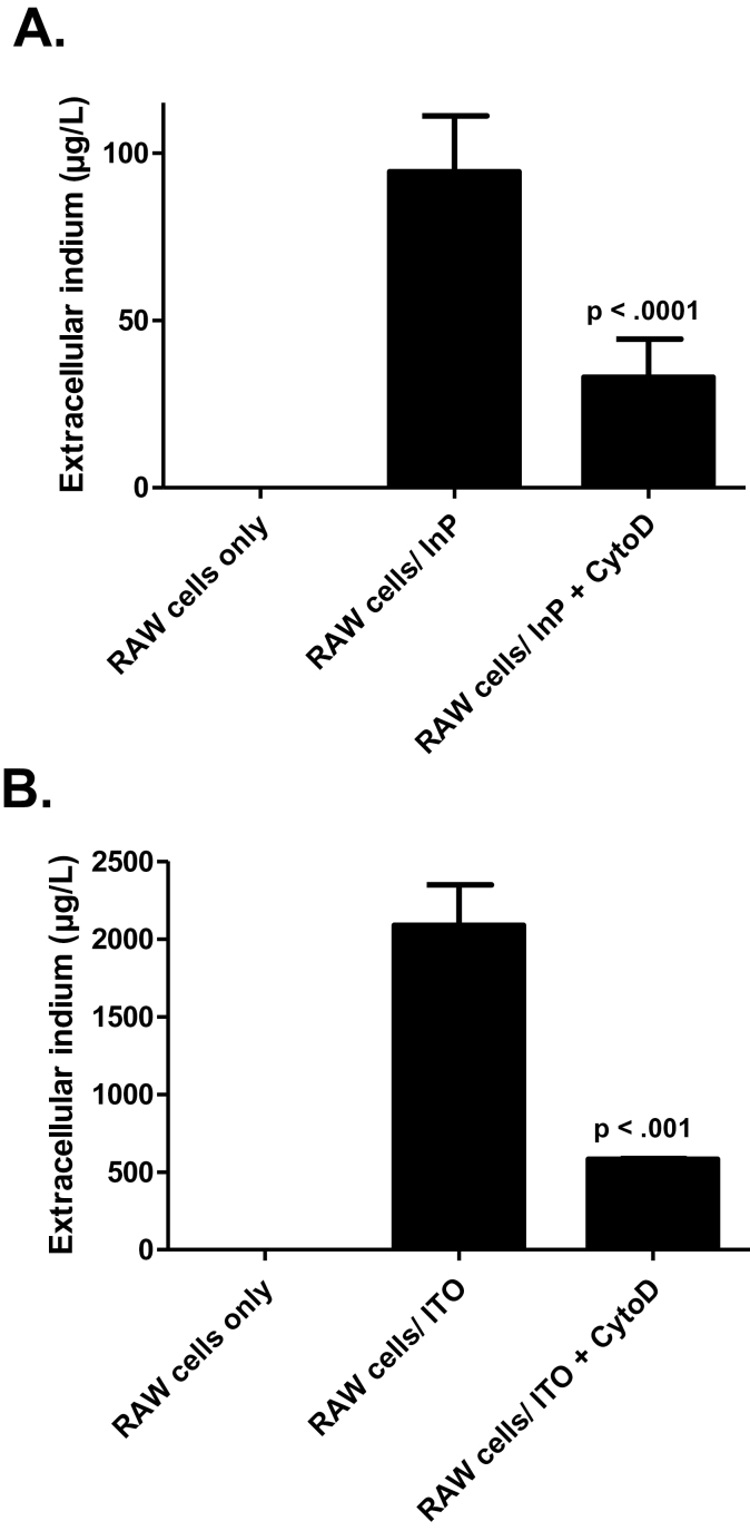
Indium-containing particles (ICPs) are used extensively in the microelectronics industry. Pulmonary toxicity is observed after inhalation exposure to ICPs; however, the mechanism(s) of pathogenesis is unclear. ICPs are insoluble at physiological pH and are initially engulfed by alveolar macrophages (and likely airway epithelial cells). We hypothesized that uptake of ICPs by macrophages followed by phagolysosomal acidification results in the solubilization of ICPs into cytotoxic indium ions. To address this, we characterized the in vitro cytotoxicity of indium phosphide (InP) or indium tin oxide (ITO) particles with macrophages (RAW cells) and lung-derived epithelial (LA-4) cells at 24h using metabolic (3-(4,5-dimethylthiazolyl-2)-2,5-diphenyltetrazolium bromide) and membrane integrity (lactate dehydrogenase) assays. InP and ITO were readily phagocytosed by RAW and LA-4 cells; however, the particles were much more cytotoxic to RAW cells and cytotoxicity was dose dependent. Treatment of RAW cells with cytochalasin D (CytoD) blocked particle phagocytosis and reduced cytotoxicity. Treatment of RAW cells with bafilomycin A1, a specific inhibitor of phagolysosomal acidification, also reduced cytotoxicity but did not block particle uptake. Based on direct indium measurements, the concentration of ionic indium was increased in culture medium from RAW but not LA-4 cells following 24-h treatment with particles. Ionic indium derived from RAW cells was significantly reduced by treatment with CytoD. These data implicate macrophage uptake and solubilization of InP and ITO via phagolysosomal acidification as requisite for particle-induced cytotoxicity and the release of indium ions. This may apply to other ICPs and strongly supports the notion that ICPs require solubilization in order to be toxic.

Keywords: ITO; InP; indium; macrophage cytotoxicity.; solubilization.


Figures

Fig. 1.
Fig. 1.
InP particles are phagocytosed by macrophages and lung-derived epithelial cells. RAW cells treated with InP (200 µg/ml) (A). RAW cells cotreated with InP + CytoD (5 µg/ml) (B). LA-4 cells treated with InP (200 µg/ml) (C). LA-4 cells cotreated with InP + CytoD (2.5 µg/ml) (D). Photomicrographs (×200 magnification) in (A and B) are RAW cells after 24h of culture and those in (C and D) are trypsinized LA-4 cells after 4h of culture.
Fig. 2.
Fig. 2.
ITO particles are phagocytosed by macrophages and lung-derived epithelial cells. RAW cells treated with ITO (400 µg/ml) (A). RAW cells cotreated with ITO + CytoD (5 µg/ml) (B). LA-4 cells treated with ITO (400 µg/ml) (C). LA-4 cells cotreated with ITO + CytoD (2.5 µg/ml) (D). Photomicrographs (×200 magnification) in (A and B) are RAW cells after 24h of culture and those in (C and D) are trypsinized LA-4 cells after 4h of culture.
Fig. 3.
Fig. 3.
Macrophages are more susceptible than lung-derived epithelial cells to the cytotoxic effects of ICPs. RAW or LA-4 cells were treated with InP or ITO particles (25, 50, 100, 200, or 400 µg/ml) or medium alone for 2h, washed once to remove extracellular particles, and then cultured for 24h in medium prior to measuring cell death by MTT (A and C) and LDH (B and D) assays. Raw absorbance values from treated cells were divided by the mean corresponding value from untreated (medium alone) control cells and expressed as fraction control (MTT) or fold over control (LDH). Data on graphs represent mean ± SD (for two to three combined experiments). RAW and LA-4 values at each dose were compared by unpaired Student’s t-test (***p < .0001, **p < .001, *p < .01, ^p < .05) Abbreviation: ns = not significant.
Fig. 4.
Fig. 4.
Cytotoxicity of ICPs for macrophages is dependent upon the phagocytic uptake of particles. RAW cells were pretreated with CytoD (5 µg/ml) or medium alone for 30min followed by treatment for 2h with medium alone, CytoD alone, InP (200 µg/ml), ITO (400 µg/ml), InP + CytoD, or ITO + CytoD. Cells were then cultured in the presence of CytoD or medium alone for 24h prior to measuring cell death by MTT (A and C) and LDH (B and D) assays. Raw absorbance values from treated cells were divided by the mean corresponding value from untreated (medium alone) control cells and expressed as fraction control (MTT) or fold over control (LDH). Data on graphs represent mean ± SD (for one representative experiment); p < .0001 is for the InP/ITO treatment group compared with the InP/ITO + CytoD and CytoD groups (one-way ANOVA with Tukey’s test).
Fig. 5.
Fig. 5.
Cytotoxicity of ICPs for macrophages is dependent upon phagolysosomal acidification. RAW cells were pretreated with BafA1 (25nM) or medium alone for 30min followed by treatment for 2h with medium alone, BafA1 alone, InP (200 µg/ml), ITO (300 µg/ml), InP + BafA1, or ITO + BafA1. Cells were then washed once to remove the BafA1 and cultured for 24h in medium prior to measuring cell death by MTT (A and C) and LDH (B and D) assays. Raw absorbance values from treated cells were divided by the mean corresponding value from untreated (medium alone) control cells and expressed as fraction control (MTT) or fold over control (LDH). Data on graphs represent mean ± SD (for one representative experiment); p < .0001 is for the InP/ITO treatment group compared with the InP/ITO + BafA1 and BafA1 groups (one-way ANOVA with Tukey’s test).
Fig. 6.
Fig. 6.
ICPs are solubilized within macrophages but not lung-derived epithelial cells, which results in the extracellular release of ionic indium. RAW or LA-4 cells were treated for 24h with InP (200 µg/ml) (A) or ITO (400 µg/ml) (C). The concentration (µg/l) of total ionic indium in cell culture medium was then measured by AAS. Control groups included supernatants from wells without cells treated with the same amount of particles in medium (background). Background indium concentration values were subtracted from the concentration values generated from particle-treated cells. LDH activity was also measured in the RAW and LA-4 cell culture medium after treatment with InP (B) or ITO (D). Data on graphs represent mean ± SD (for one representative experiment); p < .0001 is for the RAW cells + InP/ITO treatment group compared with the LA-4 cells + InP/ITO group (unpaired Student’s t-test).
Fig. 7.
Fig. 7.
ICPs are solubilized within macrophages, which is dependent upon the phagocytic uptake of particles. RAW cells were pretreated with CytoD (5 µg/ml) or medium alone for 30min followed by treatment for 24h with InP (200 µg/ml) ± CytoD (A) or ITO (400 µg/ml) ± CytoD (B). The concentration (µg/l) of total ionic indium in cell culture medium was then measured by AAS. Control groups included supernatants from wells without cells treated with the same amount of particles in medium (background). Background indium concentration values were subtracted from the concentration values generated from particle-treated cells. Data on graphs represent mean ± SD (for one representative experiment); p < .0001 in (A) is for the InP + CytoD treatment group compared with the InP group, p < .001 in (B) is for the ITO + CytoD treatment group compared with the ITO group (one-way ANOVA with Tukey’s test).

References

    1. ACGIH (2007). Threshold Limit Values for Chemical Substances and Physical Agents, and Biological Exposure Indices, p. 35 Cincinnati, OH: American Conference of Governmental Industrial Hygienists;
    1. Chibli H., Carlini L., Park S., Dimitrijevic N. M., Nadeau J. L. (2011). Cytotoxicity of InP/ZnS quantum dots related to reactive oxygen species generation. Nanoscale 3, 2552–2559 - PubMed
    1. Cho W. S., Duffin R., Howie S. E., Scotton C. J., Wallace W. A., Macnee W., Bradley M., Megson I. L., Donaldson K. (2011). Progressive severe lung injury by zinc oxide nanoparticles; the role of Zn2+ dissolution inside lysosomes. Part. Fibre Toxicol 8, 27 - PMC - PubMed
    1. Chonan T., Taguchi O., Omae K. (2007). Interstitial pulmonary disorders in indium-processing workers. Eur. Respir. J 29, 317–324 - PubMed
    1. Cummings K. J., Donat W. E., Ettensohn D. B., Roggli V. L., Ingram P., Kreiss K. (2010). Pulmonary alveolar proteinosis in workers at an indium processing facility. Am. J. Respir. Crit. Care Med 181, 458–464 - PMC - PubMed

Publication types