. 2013 Jul 30;110(31):12631-6.
doi: 10.1073/pnas.1221629110. Epub 2013 Jul 19.

Structural insights into the proton pumping by unusual proteorhodopsin from nonmarine bacteria

Affiliations

Structural insights into the proton pumping by unusual proteorhodopsin from nonmarine bacteria

Ivan Gushchin et al. Proc Natl Acad Sci U S A. .

Erratum in

  • Proc Natl Acad Sci U S A. 2013 Sep 3;110(36):14813. Arseniev, Alexander A [corrected to Arseniev, Alexander S]

Abstract

Light-driven proton pumps are present in many organisms. Here, we present a high-resolution structure of a proteorhodopsin from a permafrost bacterium, Exiguobacterium sibiricum rhodopsin (ESR). Contrary to the proton pumps of known structure, ESR possesses three unique features. First, ESR's proton donor is a lysine side chain that is situated very close to the bulk solvent. Second, the α-helical structure in the middle of the helix F is replaced by 3(10)- and π-helix-like elements that are stabilized by the Trp-154 and Asn-224 side chains. This feature is characteristic for the proteorhodopsin family of proteins. Third, the proton release region is connected to the bulk solvent by a chain of water molecules already in the ground state. Despite these peculiarities, the positions of water molecule and amino acid side chains in the immediate Schiff base vicinity are very well conserved. These features make ESR a very unusual proton pump. The presented structure sheds light on the large family of proteorhodopsins, for which structural information was not available previously.

Keywords: bacteriorhodopsin; photocycle; retinal; retinylidene protein.


Conflict of interest statement

The authors declare no conflict of interest.

Figures

Fig. 1.
Fig. 1.
Comparison of ESR with the retinylidene proteins of known structure. (A) Overlay of the known retinylidene protein structures. ESR is shown in dark blue, XR in light blue, ChR in yellow, and the other proteins in green. Most variation occurs in the helix A position. ESR differs markedly from the other proteins by disrupted α-helical structure in the middle part of the helix F. The helices A, E, F, and G are labeled. N-terminal residues of ChR are not shown. (B) π-bulge and 310-helix–like structures in the ESR’s helix F. The disruption of normal α-helical structure is stabilized by the hydrogen bonds between N224 and the carbonyl oxygen of G190, and between W154 and the carbonyl oxygen of I187. Whereas the proline 195 is conserved in other proteins, disruption of α-helical structure in the region of residues 187–191 is unique to ESR. (C) Phylogenetic tree showing the relations between the retinylidene proteins of known structure, ESR, and other proteorhodopsins. Residues W154 and N224 are unique to the proteorhodopsin family. Lysine at the proton donor position is also observed in the clade B proteorhodopsins (genes pop and pop-1). The multiple sequence alignment and the phylogenetic tree were generated using ClustalX 2.1 (39), and the tree was drawn using the TreeDyn web server (40). The proteins used for comparison are channelrhodopsin (ChR), PDB ID code 3UG9 (41); bacteriorhodopsin (BR), PDB ID code 1C3W (22); archaerhodopsins 1 and 2 (aR-1 and aR-2), PDB ID codes 1UAZ and 1VGO (42); H. salinarum halorhodopsin (HsHR), PDB ID code 1E12 (43); Natronomonas pharaonis halorhodopsin (NpHR), PDB ID code 3A7K (44); N. pharaonis sensory rhodopsin II (NpSRII), PDB ID code 3QAP (45); Anabaena sensory rhodopsin (ASR), PDB ID code 1XIO (46); Acetabularia rhodopsin 2 (AR2), PDB ID code 3:00 AM6 (47); and xanthorhodopsin (XR), PDB ID code 3DDL (12).
Fig. 2.
Fig. 2.
Structure of the ESR Schiff base-proximal region with electron density maps. (A) View along the retinal. (B) View perpendicular to the retinal. Aspartates 85 and 221 are traditionally close to the Schiff base, with a water molecule (W402) in between. There is a histidine residue (H57) close to D85. There are also additional electron densities that can be treated as partially ordered water molecules (W406 and W407). The arginine 82 side chain points away from the Schiff base and forms hydrogen bonds with E130.
Fig. 3.
Fig. 3.
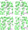
Comparison of the ESR, XR, and BR retinal binding pockets in the ground state. Both ESR and XR differ from BR by presence of the histidine residue not far from the retinal. ESR's retinal pocket also possesses other differences. First and probably most important, its histidine is turned toward the arginine 82, which in its turn is removed from the retinal, similarly to the bacteriorhodopsin's M state. Second, in ESR, the residue preceding the retinal-bound lysine is asparagine, as opposed to alanine in BR, XR, and most of other proton-pumping bacterial rhodopsins.
Fig. 4.
Fig. 4.
Comparison of the ESR, XR, and BR proton uptake regions. The conformations differ in the two observed ESR molecules. Contrary to BR, XR, and other light-driven proton pumps of known structure, ESR has a lysine (Lys-96) residue at the proton uptake, whose electron densities are shown at the level of 1.4σ. The cavity around Lys-96 is mostly surrounded by hydrophobic amino acids and may accommodate at least three water molecules. The only polar residue, Thr-43, separates the cavity from the bulk solvent. On the contrary, the proton uptake residues Glu-107 of XR and Asp-96 of BR are far from the bulk solvent, being separated from it by Ser-48 and Tyr-45 in XR and Phe-42 in BR. Lys-96 side chain is ordered in one ESR molecule and partially disordered in the other. Additional positive electron densities around Lys-96 (not shown) are observed in the difference maps that are probably related to mobile water molecules.
Fig. 5.
Fig. 5.
The cavities on the putative proton path in ESR, XR, and BR that may be occupied by water molecules. The upmost and the lowest cavities on the figure face the bulk solvent. The important ionizable and charged residues are shown explicitly. In the proton uptake region, ESR’s Lys-96 is separated only by the Thr-43 side chain from the solvent whereas there is a large gap in XR and BR between the bulk and Glu-107 and Asp-96 correspondingly. There is enough space for at least three water molecules around Lys-96 in ESR. In ESR, the histidine residue of a putative proton release group Asp-221/His-57 is immersed in a cavity of a size sufficient for a water molecule from the bulk to come in contact with it. This continuous cavity contains the ordered water molecules 402 and 406, and transitions into the bulk. In XR, the release group is shielded by Arg-93. In BR, the proton is released from the completely different group, a pair of glutamates (Glu-194 and Glu-204) that are separated by the Ser-193 side chain from the bulk. The cavities are determined as a composition of the crystallographically recognizable water molecules and the space determined using the Hollow software (48) with a 1.4-Å probe and 0.2-Å grid spacing.
Fig. 6.
Fig. 6.
The models of the BR and ESR photocycles (11, 27). Some of the transitions are reversible. The timescales are very approximate as they depend strongly on the conditions such as pH, temperature, and other factors.

References

    1. Spudich JL, Yang C-S, Jung K-H, Spudich EN. Retinylidene proteins: Structures and functions from archaea to humans. Annu Rev Cell Dev Biol. 2000;16:365–392. - PubMed
    1. Pastrana E. Optogenetics: Controlling cell function with light. Nat Methods. 2011;8:24–25.
    1. Hirai T, Subramaniam S, Lanyi JK. Structural snapshots of conformational changes in a seven-helix membrane protein: Lessons from bacteriorhodopsin. Curr Opin Struct Biol. 2009;19(4):433–439. - PMC - PubMed
    1. Oesterhelt D, Stoeckenius W. Rhodopsin-like protein from the purple membrane of Halobacterium halobium. Nat New Biol. 1971;233(39):149–152. - PubMed
    1. Béjà O, et al. Bacterial rhodopsin: Evidence for a new type of phototrophy in the sea. Science. 2000;289(5486):1902–1906. - PubMed

Publication types

MeSH terms

Associated data