

Prolactin cooperates with loss of p53 to promote claudin-low mammary carcinomas
- PMID: 23873024
- PMCID: PMC4007359
- DOI: 10.1038/onc.2013.278
Prolactin cooperates with loss of p53 to promote claudin-low mammary carcinomas
Abstract
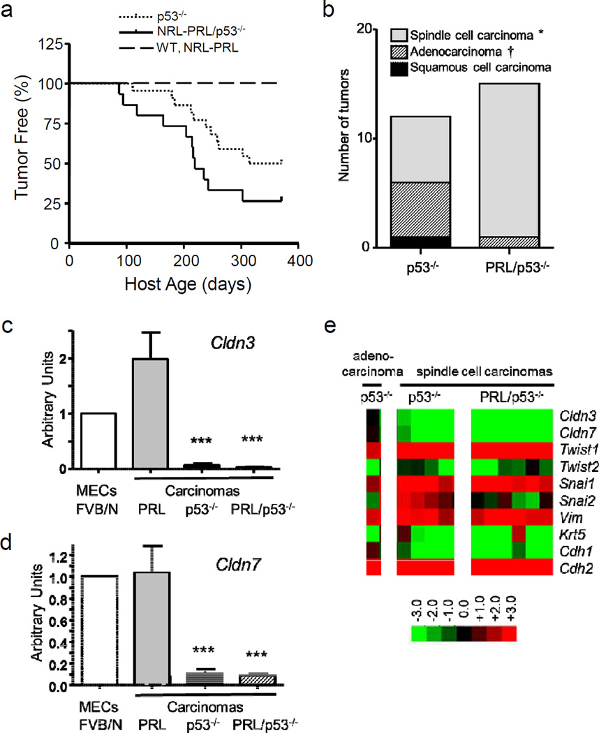
TP53 is one of the most commonly mutated genes in cancer. In breast cancer, it is mutated in about 40% of primary clinical tumors and is associated with poor survival. The mammotrophic hormone, prolactin (PRL), and/or its receptor are also expressed in many breast cancers, and accumulating epidemiologic data link PRL to breast cancer development and progression. Like TP53 mutations, evidence for PRL activity is evident across several molecular cancer subtypes, and elevated PRL expression and loss of p53 have been observed in some of the same clinical tumors. In order to examine the interaction of these factors, we used genetically modified mouse models of mammary-specific p53 loss and local overexpression of PRL. We demonstrated that mammary PRL decreased the latency of tumors in the absence of p53, and increased the proportion of triple-negative claudin-low carcinomas, which display similarities to human clinical metaplastic carcinomas. Moreover, PRL/p53(-/-) carcinomas displayed higher rates of proliferation and more aggressive behavior. Transcripts associated with cell cycle progression, invasion and stromal reactivity were differentially expressed in carcinomas that developed in the presence of elevated PRL. PRL/p53(-/-) carcinomas also exhibited selectively altered expression of activating protein-1 components, including higher levels of c-Jun and FosL1, which can drive transcription of many of these genes and the epithelial-mesenchymal transition. The ability of PRL to promote claudin-low carcinomas demonstrates that PRL can influence this subset of triple-negative breast cancers, which may have been obscured by the relative infrequency of this cancer subtype. Our findings suggest novel therapeutic approaches, and provide a preclinical model to develop possible agents.
Conflict of interest statement
The authors have nothing to disclose.
Figures


Similar articles
-
Prolactin-induced mouse mammary carcinomas model estrogen resistant luminal breast cancer.Breast Cancer Res. 2011 Jan 28;13(1):R11. doi: 10.1186/bcr2819. Breast Cancer Res. 2011. PMID: 21276249 Free PMC article.
-
Met synergizes with p53 loss to induce mammary tumors that possess features of claudin-low breast cancer.Proc Natl Acad Sci U S A. 2013 Apr 2;110(14):E1301-10. doi: 10.1073/pnas.1210353110. Epub 2013 Mar 18. Proc Natl Acad Sci U S A. 2013. PMID: 23509284 Free PMC article.
-
Ovarian hormones are not required for PRL-induced mammary tumorigenesis, but estrogen enhances neoplastic processes.J Endocrinol. 2009 Oct;203(1):99-110. doi: 10.1677/JOE-09-0221. Epub 2009 Jul 27. J Endocrinol. 2009. PMID: 19635758 Free PMC article.
-
The C3(1)/SV40 T-antigen transgenic mouse model of mammary cancer: ductal epithelial cell targeting with multistage progression to carcinoma.Oncogene. 2000 Feb 21;19(8):1020-7. doi: 10.1038/sj.onc.1203280. Oncogene. 2000. PMID: 10713685 Review.
-
The role of prolactin and growth hormone in breast cancer.Oncogene. 2000 Feb 21;19(8):1072-6. doi: 10.1038/sj.onc.1203349. Oncogene. 2000. PMID: 10713692 Review.
Cited by
-
Modeling prolactin actions in breast cancer in vivo: insights from the NRL-PRL mouse.Adv Exp Med Biol. 2015;846:201-20. doi: 10.1007/978-3-319-12114-7_9. Adv Exp Med Biol. 2015. PMID: 25472540 Free PMC article.
-
Suppression of Breast Cancer by Small Molecules That Block the Prolactin Receptor.Cancers (Basel). 2021 May 28;13(11):2662. doi: 10.3390/cancers13112662. Cancers (Basel). 2021. PMID: 34071395 Free PMC article.
-
Emerging cytokine networks in colorectal cancer.Nat Rev Immunol. 2015 Oct;15(10):615-29. doi: 10.1038/nri3896. Epub 2015 Sep 11. Nat Rev Immunol. 2015. PMID: 26358393 Review.
-
Prolactin: The Third Hormone in Breast Cancer.Front Endocrinol (Lausanne). 2022 Jun 16;13:910978. doi: 10.3389/fendo.2022.910978. eCollection 2022. Front Endocrinol (Lausanne). 2022. PMID: 35784527 Free PMC article. Review.
-
Antiestrogen Therapy Increases Plasticity and Cancer Stemness of Prolactin-Induced ERα+ Mammary Carcinomas.Cancer Res. 2018 Apr 1;78(7):1672-1684. doi: 10.1158/0008-5472.CAN-17-0985. Epub 2018 Jan 23. Cancer Res. 2018. PMID: 29363543 Free PMC article.
References
-
- Sorlie T, Perou CM, Tibshirani R, Aas T, Geisler S, Johnsen H, Hastie T, Eisen MB, van de Rijn M, Jeffrey SS, Thorsen T, Quist H, Matese JC, Brown PO, Botstein D, Lonning PE, Borresen-Dale AL. Gene expression patterns of breast carcinomas distinguish tumor subclasses with clinical implications. Proc Natl Acad Sci U S A. 2001;98:10869–10874. - PMC - PubMed
-
- Jackson JG, Lozano G. The mutant p53 mouse as a pre-clinical model. Oncogene. 2013 - PubMed
-
- Jerry DJ, Kittrell FS, Kuperwasser C, Laucirica R, Dickinson ES, Bonilla PJ, Butel JS, Medina D. A mammary-specific model demonstrates the role of the p53 tumor suppressor gene in tumor development. Oncogene. 2000;19:1052–1058. - PubMed
Publication types
MeSH terms
Substances
Grants and funding
LinkOut - more resources
Full Text Sources
Other Literature Sources
Molecular Biology Databases
Research Materials
Miscellaneous
