. 2013 Aug 29;500(7464):589-92.
doi: 10.1038/nature12358. Epub 2013 Jul 21.

Coordination of heart and lung co-development by a multipotent cardiopulmonary progenitor

Affiliations

Coordination of heart and lung co-development by a multipotent cardiopulmonary progenitor

Tien Peng et al. Nature. .

Abstract

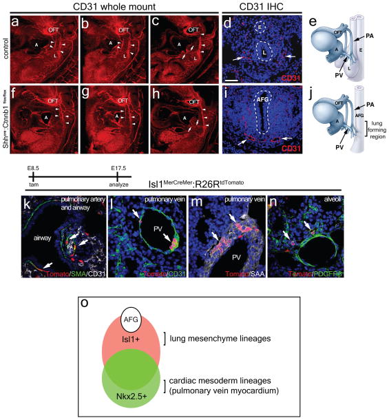
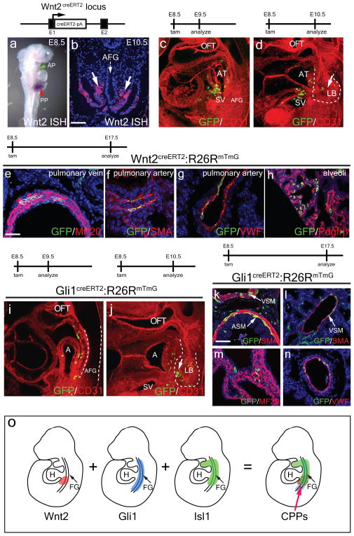
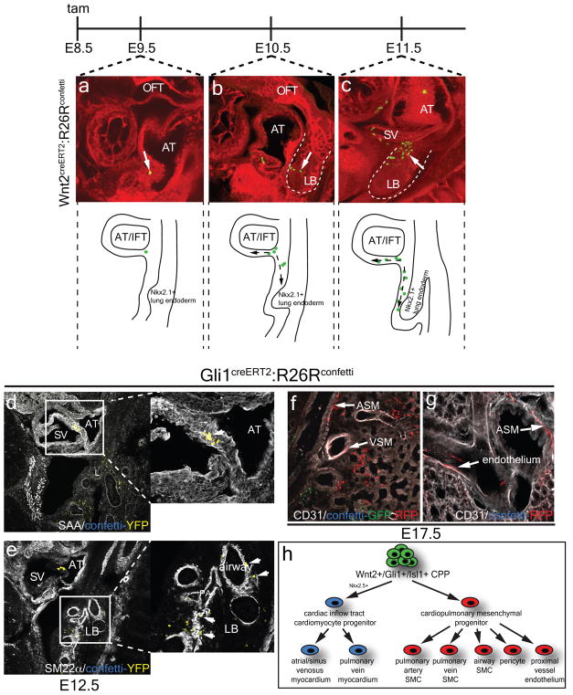
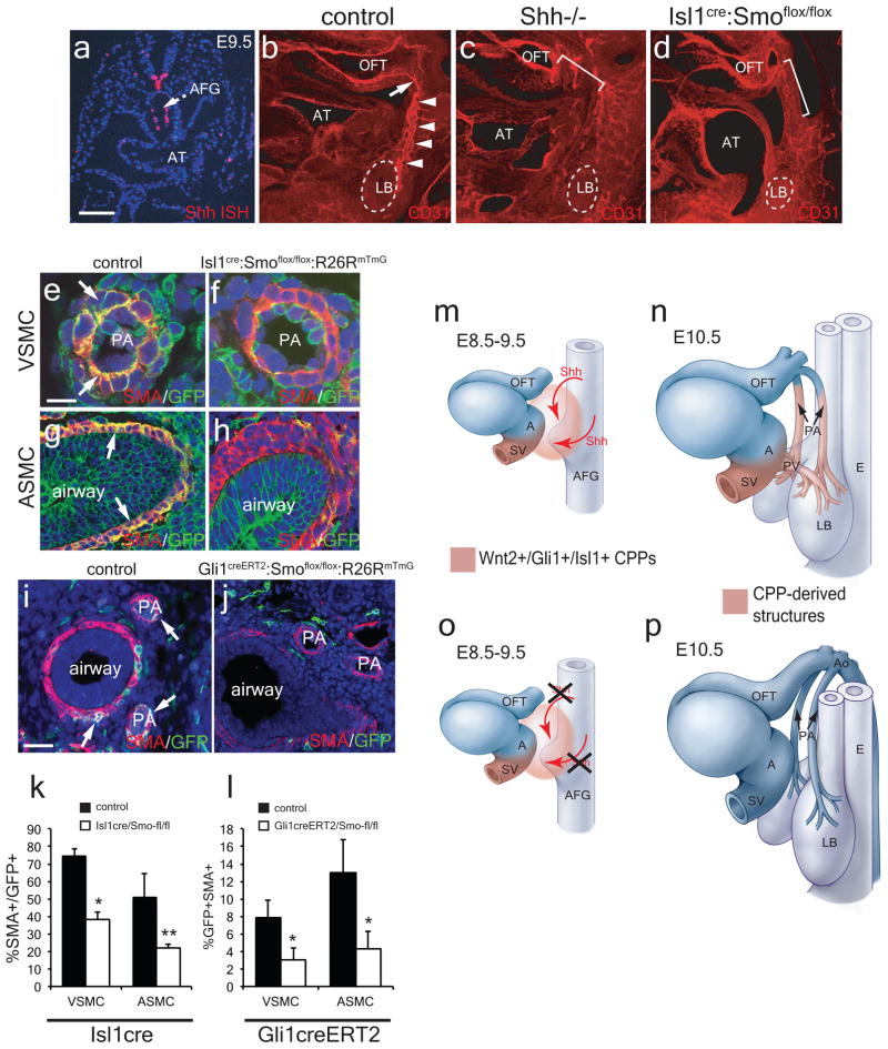
Co-development of the cardiovascular and pulmonary systems is a recent evolutionary adaption to terrestrial life that couples cardiac output with the gas exchange function of the lung. Here we show that the murine pulmonary vasculature develops even in the absence of lung development. We have identified a population of multipotent cardiopulmonary mesoderm progenitors (CPPs) within the posterior pole of the heart that are marked by the expression of Wnt2, Gli1 and Isl1. We show that CPPs arise from cardiac progenitors before lung development. Lineage tracing and clonal analysis demonstrates that CPPs generate the mesoderm lineages within the cardiac inflow tract and lung including cardiomyocytes, pulmonary vascular and airway smooth muscle, proximal vascular endothelium, and pericyte-like cells. CPPs are regulated by hedgehog expression from the foregut endoderm, which is required for connection of the pulmonary vasculature to the heart. Together, these studies identify a novel population of multipotent cardiopulmonary progenitors that coordinates heart and lung co-development that is required for adaptation to terrestrial existence.


Conflict of interest statement

Competing financial interests

The authors declare no competing financial interests

Figures

Figure 1
Figure 1. The pulmonary vasculature develops in the absence of lung specification
The paired PAs are observed tracking in a posterior direction in both the control (a–c, arrowheads) and Shhcre:Ctnnb1flox/flox mutants (f–h, arrowheads) at E10.5 using CD31 whole mount immunostaining and section immunostaining (d and i, arrows). The primitive PVs are observed emerging from the atria and extending towards the region of the foregut where the lung would normally form in both control and Shhcre:Ctnnb1flox/flox mutants (c and h, arrows). These developmental hallmarks are illustrated in models (e and j). Isl1+ progenitors tagged at E8.5 give rise to the PA and airway smooth muscle (k), endothelial (l), myocardial (m), and Pdgfrβ+ pericyte-like (n) lineages of the pulmonary vasculature and other mesodermal derivatives. This overlapping but distinct origin of lung mesoderm derivatives is diagrammed in (o). PA=pulmonary artery, PV=pulmonary vein, AT=atria, AFG=anterior foregut. OFT=outflow tract, L=lung, T=trachea, E=esophagus. Control for a–h= Shhcre:Ctnnb1flox/+. Scale bar (g and h)=100 μm.
Figure 2
Figure 2. Wnt2+ and Gli1+ cells define a cardiopulmonary progenitor (CPP) and generate mesoderm derivatives of the lung and cardiac inflow tract
Wnt2 is expressed in the ventral mesoderm surrounding the anterior foregut and overlapping the posterior pole of the heart (a and b, arrows). Wnt2+ cells tagged at E8.5 can generate cells within the cardiac inflow tract as well as in the lung mesoderm (c and d). Wnt2+ cells tagged at E8.5 generate myocardium of the PV, smooth muscle of the PA, endothelium of the proximal PA, and Pdgfrβ+ pericyte-like cells in the lung by E17.5 (e–h). Gli1+ cells generate derivatives within the inflow tract and early lung mesoderm similar to Wnt2+ cells (i and j) and generate mesoderm lineages within the lung including airway and vascular smooth muscle (k and l), and endothelium of the proximal pulmonary vessels (n) as well as the myocardium of the atria (m). CPPs are located in a region of overlapping Wnt2, Gli1, and Isl1 expression between the developing heart and the anterior foregut (o). AP=anterior pole of the heart, PP=posterior pole of the heart, AFG=anterior foregut, AT=atria, OFT=outflow tract, PA=pulmonary artery, PV=pulmonary vein, LB=lung bud, ASM=airway smooth muscle, VSM=vascular smooth muscle, SMA=smooth muscle actin, SAA= sarcomeric α-actinin, VWF=von Willibrand factor. Scale bars b=100 μm, e–h=50 μm, k–n=50 μm.
Figure 3
Figure 3. Clonal analysis reveals that CPPs generate related lineages within the cardiopulmonary system
Single clones of Wnt2+ CPPs contribute to both the developing cardiac inflow tract as well as the mesoderm of the lung (a–c). Clonal analysis with the Gli1creERT2:R26Rconfetti line shows that a single CPP clone (YFP+) can generate myocardium (SAA+) within the cardiac inflow tract (d, arrow) as well as lung mesodermal lineages such as smooth muscle (SM22α+) around the airway and PA (e, arrows). Clonal analysis of Gli1creERT2:R26Rconfetti lungs show that vascular and airway smooth muscle and endothelium of the proximal vessels share a common Gli1+ progenitor at E8.5 (f and g). A cell lineage tree showing the relationship of CPPs to differentiated lineages within the cardiopulmonary system (h). AT=atria, OFT=outflow tract, LB=lung bud, SV=sinus venosus, ASM=airway smooth muscle, VSM=vascular smooth muscle, SAA= sarcomeric α-actinin.
Figure 4
Figure 4. Hedgehog signaling is required in CPPs to coordinate the vascular connection between the heart and lung
Shh expression at E9.5 in the anterior foregut endoderm by in situ hybridization (a). Shh−/− mutants and Isl1cre:Smoflox/flox mutants display a disrupted vascular plexus between the developing heart and lung at E10.5 (b–d, brackets, control=Shh +/−). Inactivation of Smo within Isl1+ CPPs inhibits their contribution to airway and vascular smooth muscle at E13.5 (e–h, k, control= Isl1cre:Smoflox/+:R26RmTmG). Inactivation of Smo within Gli1+ CPPs inhibits their contribution to airway and vascular smooth muscle at E13.5 (i, j, l, control= Gli1creERT2:Smoflox/+:R26RmTmG). CPPs orchestrate lung and cardiac co-development and are regulated in turn by Shh expression from the anterior foregut endoderm (m–p). AT=atria, AFG=anterior foregut, OFT=outflow tract, LB=lung bud, PA=pulmonary artery, Ao=aorta, PV=pulmonary vein, SV=sinus venosus, SVC and IVC=superior and inferior vena cava. *p<0.01, **p<0.05. Error bars=S.D. Scale bars A=50 μm, E-h=20 μm, i and j=50 μm.

Similar articles

Cited by

References

    1. Peng T, Morrisey EE. Development of the pulmonary vasculature: Current understanding and concepts for the future. Pulm Circ. 2013;3:176–178. doi: 10.4103/2045-8932.109954. - DOI - PMC - PubMed
    1. Goss AM, et al. Wnt2/2b and beta-catenin signaling are necessary and sufficient to specify lung progenitors in the foregut. Dev Cell. 2009;17:290–298. doi: 10.1016/j.devcel.2009.06.005. - DOI - PMC - PubMed
    1. Harris-Johnson KS, Domyan ET, Vezina CM, Sun X. beta-Catenin promotes respiratory progenitor identity in mouse foregut. Proc Natl Acad Sci U S A. 2009;106:16287–16292. doi: 10.1073/pnas.0902274106. - DOI - PMC - PubMed
    1. Cai CL, et al. Isl1 identifies a cardiac progenitor population that proliferates prior to differentiation and contributes a majority of cells to the heart. Dev Cell. 2003;5:877–889. - PMC - PubMed
    1. Moses KA, DeMayo F, Braun RM, Reecy JL, Schwartz RJ. Embryonic expression of an Nkx2-5/Cre gene using ROSA26 reporter mice. Genesis. 2001;31:176–180. - PubMed

Publication types

MeSH terms