. 2013:4:2183.
doi: 10.1038/ncomms3183.

Small molecules enable neurogenin 2 to efficiently convert human fibroblasts into cholinergic neurons

Affiliations

Small molecules enable neurogenin 2 to efficiently convert human fibroblasts into cholinergic neurons

Meng-Lu Liu et al. Nat Commun. 2013.

Abstract

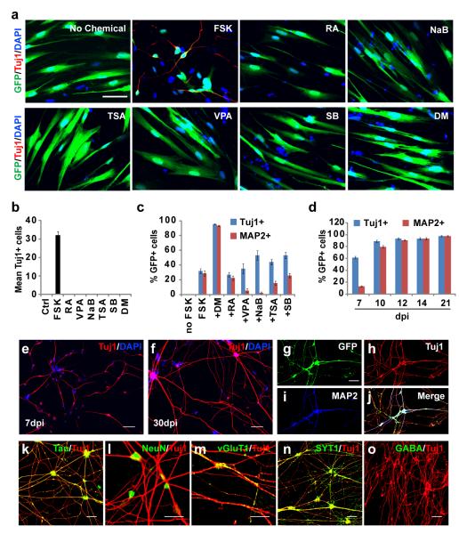
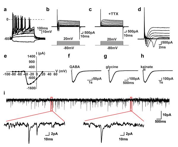
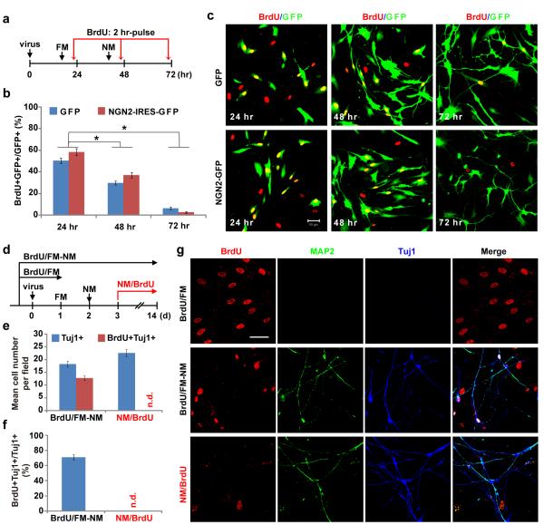
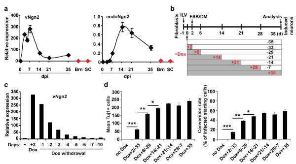
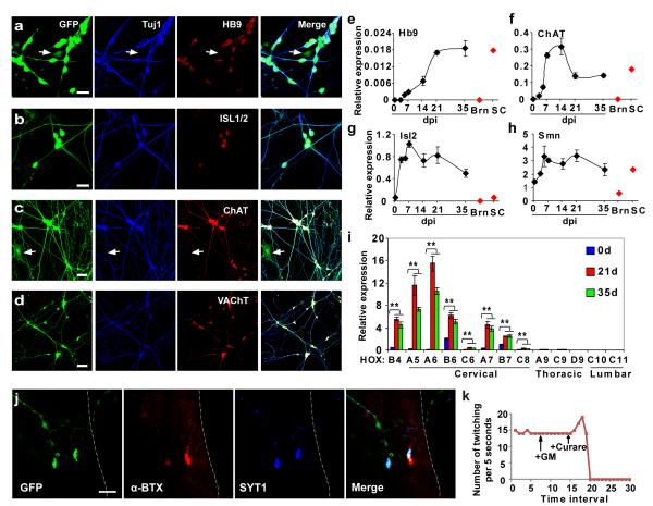
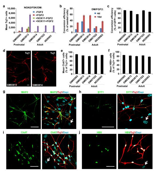
Cell fate can be reprogrammed by modifying intrinsic and extrinsic cues. Here we show that two small molecules (forskolin and dorsomorphin) enable the transcription factor Neurogenin 2 (NGN2) to convert human fetal lung fibroblasts into cholinergic neurons with high purity (>90%) and efficiency (up to 99% of NGN2-expressing cells). The conversion is direct without passing through a proliferative progenitor state. These human induced cholinergic neurons (hiCN) show mature electrophysiological properties and exhibit motor neuron-like features, including morphology, gene expression and the formation of functional neuromuscular junctions. Inclusion of an additional transcription factor, SOX11, also efficiently converts postnatal and adult skin fibroblasts from healthy and diseased human patients to cholinergic neurons. Taken together, this study identifies a simple and highly efficient strategy for reprogramming human fibroblasts to subtype-specific neurons. These findings offer a unique venue for investigating the molecular mechanisms underlying cellular plasticity and human neurodegenerative diseases.


Figures

Figure 1
Figure 1. NGN2 induces rapid and efficient neuronal conversion of human fetal fibroblasts
(a) FSK but not any other tested small molecules synergizes with NGN2 to generate Tuj1+ cells from IMR-90 fetal fibroblasts. GFP indicates cells transduced with NGN2-expressing virus. FSK, forskolin; RA, retinoic acid; VPA, valproic acid; NaB, sodium n-butyric acid; TSA, trichostatin A; SB, SB431542; DM, dorsomorphin. (b) Quantification of Tuj1+ cells 5 days post chemical treatments (means ± s.e.m., n=40 randomly selected 20x fields from triplicate samples). (c) DM promotes conversion efficiency, neuronal survival and maturation. Cells were counted 10 days post treatment (means ± s.e.m., n=30 random 20x fields from triplicate samples). (d) Time-dependent maturation of converted neurons by NGN2/FSK/DM (means ± s.e.m., n=10 random 20x fields from triplicate samples). (e-f) Representative morphology of induced neurons at 7 or 30 dpi. (g-l) Converted cells express markers for mature neurons. (m-n) Induced neurons express glutamatergic (vGlut1) and presynaptic (synaptotagmin 1, SYT1) markers, which showed a punctate staining pattern. (o) GABA is rarely detected in converted neurons. Scales, 50 μm for (a, e-k, n-o), 20 μm for (l, m).
Figure 2
Figure 2. NGN2-induced neurons are functionally mature
(a) Fibroblast-derived neurons fire repetitive action potentials by depolarizing current injections (n=10/10 recorded cells). (b-c) TTX-sensitive fast inward currents upon depolarization (n=7/7 recorded cells). (d-e) Sodium currents elicited at or above −30 mV, with a peak current at −1200 pA. (f-h) Inward currents induced by puffing inhibitory or excitatory neurotransmitters (n=3/4 for GABA, n=3/5 for glycine, and n=3/3 for kainate). (i) Induced neurons exhibit spontaneous activity indicating synaptic connections between neurons (n=19/19 of recorded cells).
Figure 3
Figure 3. Neuronal conversion from human fibroblasts is accompanied by cell cycle exit
(a) Experimental design for data in (b-c). Cells were incubated with BrdU for 2 hr before immunocytochemistry. FM, fibroblast medium; NM, neuronal induction medium containing FSK and DM. (b-c) Treatment with neuronal induction medium quickly stopped proliferation of cells transduced with either GFP or NGN2-IRES-GFP viruses (means ± s.e.m., n=15 randomly selected 20x fields from triplicate samples, *p < 0.001 by Student’s t-test). Scale, 50 μm. (d) Experimental design for data in (e-g). Cells were treated with BrdU for the indicated periods of time. (e-g) A majority of the cells were labeled with BrdU if treated prior to neuronal conversion (BrdU/FM-NM), whereas cells were not labeled by BrdU if treated one day after switching to neuronal induction medium (NM/BrdU). Quantitative data are means ± s.e.m. from 20 randomly selected 20x fields from triplicate samples. n.d., not detected. Scale, 50 μm.
Figure 4
Figure 4. Neuronal conversion from human fibroblasts does not require long-term expression of NGN2
(a) qRT-PCR analysis of retrovirus-expressed NGN2 (vNGN2) and the induction of endogenous NGN2 (endoNGN2) at the indicated time points after retroviral transduction of human fetal fibroblasts (means ± s.e.m., n=3 at each time point). Samples from normal adult human brain (Brn) and spinal cord (SC) were used as controls (n=1). (b) Experimental design using doxycycline (Dox) inducible expression system. Cells (5,000/well in 48-well plate) were cotransduced with lentivirus constitutively expressing rtTA3G and Dox-inducible lentivirus(iLV) expressing NGN2-IRES-GFP under the TRE3G promoter. One day post transduction, Dox (0.1 μg/ml) was applied for the indicated periods of time and then withdrawn. The transduction efficiency was about 8% indicated by initial GFP expression. Tuj1-expression was analyzed 35 days later. (c) qRT-PCR analysis of exogenous NGN2 expression, which returns to basal level 6 days after Dox withdrawal (n=1). (d) NGN2 expression is no longer required after neuronal conversion at 21 dpi, although it increases total number of converted neurons at earlier time points. Tuj1+ cells were quantified from a whole well from triplicate samples (means ± s.e.m., n=3). *p=0.04, **p=0.003, and ***p=0.0003 by Student’s t-test.
Figure 5
Figure 5. Cholinergic motor neuron-like cells converted from human fetal fibroblasts
(a-d) Immunocytochemistry analysis of induced neurons with markers for cholinergic motor neurons, including HB9 (91.7%, n=325), ISL1/2 (90.2%, n=347), ChAT (98.5%, n=325), and VAChT (98.2%, n=273). GFP expression indicates NGN2-transduced cells. Control virus-transduced fibroblasts were stained negative for each of these markers. Arrows indicate cells stained negative for HB9 or ChAT. (e-i) Gene expression analysis by qRT-PCR during the reprogramming process (means ± s.e.m., n=3 at each time point). It was normalized to that of house-keeping gene Hprt. Samples from normal adult human brain (Brn) and spinal cord (SC) were used as controls (n=1). **p < 0.01 by Student’s t-test. (j) Immunocytochemistry analysis of synaptic connections between hiCNs and myotubes in co-culture. A multinucleated myotube is outlined. α-BTX, α-bungarotoxin; SYT1, synaptotagmin 1. (k) Quantification of rhythmic contractions of myotubes after co-culturing with hiCNs. Such contractions can be blocked by curare, a competitive inhibitor of acetylcholine receptor (n=5 independent myotubes). Scales, 20 μm for (a-b), 50 μm for (c-d), and 10 μm for (j).
Figure 6
Figure 6. Efficient generation of cholinergic neurons from postnatal and adult human skin fibroblasts
(a) SOX11 and FGF2 facilitate NGN2 to convert postnatal (GM05565 and GM03813) or adult (GM03814, GM07522, and ND29563) human skin fibroblasts to neurons. Tuj1+ cells were quantified from triplicate samples at 21 dpi (means ± s.e.m.). (b-c) Conversion efficiency normalized to total number of seeded cells (b) or NGN2-expressing cells (c). The total number of Tuj1+ cells from triplicate samples was quantified at 21 dpi (means ± s.e.m.). A longer treatment of DM/FGF2 enhances conversion efficiency. (d) Representative images of converted Tuj1+ neurons from postnatal (GM03813) and adult (GM03814, the mother of GM03813) fibroblasts at 21 dpi. (e-f) Quantification of converted neurons expressing ChAT (e, 81.9%-90.7%) or HB9 (f, 78.0%-85.9%). The total number of Tuj1+, ChAT+, or HB9+ cells from triplicate samples was quantified at 21 dpi (means ± s.e.m.). (g-j) Induced neurons from skin fibroblasts (GM07522) express pan-neuronal marker MAP2 (g) and presynaptic marker SYT1 (h). (i-j) Converted neurons from skin fibroblasts (GM03813) are cholinergic (ChAT+, i) and express markers for early motor neurons (HB9+, j). Arrows show cells stained negative for the indicated neuronal markers. Scales, 50 μm.

Similar articles

Cited by

References

    1. Hanna JH, Saha K, Jaenisch R. Pluripotency and cellular reprogramming: facts, hypotheses, unresolved issues. Cell. 2010;143:508–525. - PMC - PubMed
    1. Ladewig J, et al. Small molecules enable highly efficient neuronal conversion of human fibroblasts. Nat. Methods. 2012;9:575–578. - PubMed
    1. Marro S, et al. Direct lineage conversion of terminally differentiated hepatocytes to functional neurons. Cell Stem Cell. 2011;9:374–382. - PMC - PubMed
    1. Son EY, et al. Conversion of mouse and human fibroblasts into functional spinal motor neurons. Cell Stem Cell. 2011;9:205–218. - PMC - PubMed
    1. Caiazzo M, et al. Direct generation of functional dopaminergic neurons from mouse and human fibroblasts. Nature. 2011;476:224–227. - PubMed

Publication types

MeSH terms

Associated data