. 2013 Dec 1;521(17):3954-71.
doi: 10.1002/cne.23386.

Thalamic input to representations of the teeth, tongue, and face in somatosensory area 3b of macaque monkeys

Affiliations

Thalamic input to representations of the teeth, tongue, and face in somatosensory area 3b of macaque monkeys

Christina M Cerkevich et al. J Comp Neurol. .

Abstract

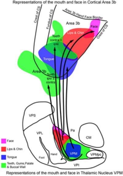
Representations of the parts of the oral cavity and face in somatosensory area 3b of macaque monkeys were identified with microelectrode recordings and injected with different neuroanatomical tracers to reveal patterns of thalamic projections to tongue, teeth, and other representations in primary somatosensory cortex. The locations of injection sites and resulting labeled neurons were further determined by relating sections processed to reveal tracers to those processed for myeloarchitecture in the cortex and multiple architectural stains in the thalamus. The ventroposterior medial subnucleus (VPM) for touch was identified as separate from the ventroposterior medial parvicellular nucleus (VPMpc) for taste by differential expression of several types of proteins. Our results revealed somatotopically matched projections from VPM to the part of 3b representing intra-oral structures and the face. Retrogradely labeled cells resulting from injections in area 3b were also found in other thalamic nuclei including: anterior pulvinar (Pa), ventroposterior inferior (VPI), ventroposterior superior (VPS), ventroposterior lateral (VPL), ventral lateral (VL), center median (CM), central lateral (CL), and medial dorsal (MD). None of our injections, including those into the representation of the tongue, labeled neurons in VPMpc, the thalamic taste nucleus. Thus, area 3b does not appear to be involved in processing taste information from the thalamus. This result stands in contrast to those reported for New World monkeys.

Keywords: gustatory thalamus; somatosensory cortex; ventroposterior thalamus.


Conflict of interest statement

Conflict of Interest: The authors have no conflict of interest.

Figures

Figure 1
Figure 1. Histological characteristics of the somatosensory thalamus
Photomicrographs of coronal sections of thalamus stained for CO (column A) and Cb (column B) cut at 40 µm thickness. Representative sections were selected to show the borders of nuclei pertinent to cell labeling from the injections of tracers, including the somatosensory VPM and the gustatory VPMpc. There is approximately 1.16 mm between rostral (CO 137 and Cb 135) and caudal (CO 107 and Cb 105) sections. Cb sections are the closet to the CO sections. Abbreviations as in table 1. Scale bar 2 mm.
Figure 2
Figure 2. Comparison of CO and VGluT2 staining in the thalamus
(A-B) Photomicrographs of coronal thalamus sections stained for CO (A) or VGluT2 (B). Black lines mark anatomical borders and CO or VGluT2 dense patches within VPM. Circles indicate blood vessels and other landmarks. (C) Drawings of aligned borders from these CO and VGluT2 sections. Black lines mark borders of thalamic nuclei revealed by CO staining. CO densities in VPM are shaded in gray. Gray lines indicate borders of thalamic nuclei revealed by VGluT2 expression. VGluT2 patches in VPM are outlined in dark gray. CO densities have been shown to represent different parts of the face and mouth (Rausell & Jones, 1991a). When aligned according to landmarks, VGluT2 dense patches in VPM often subdivided larger CO dense patches in the structure. These smaller VGluT2 dense subdivisions of CO patches likely represent an even more fine-grained anatomical representation of the parts face and mouth within VPM. There is a slight mismatch between dense patches of CO and VGluT2 expression because the two sections are not immediately adjacent, with 80 µm between them. Intervening sections were used for staining to reveal tracers and different immunohistochemical stains. Coronal sections are 40 µm thick. Abbreviations as in table 1. Scale bar is 1mm.
Figure 3
Figure 3. Locations of injection sites in the central sulcus and the injected representations
A-D. Markers in the open central sulcus indicate the center of each tracer injection in the representations indicated below. Area 3b is shaded in gray. The somatotopic organization of area 3b from mouth (lateral) to foot (medial) is outlined, adapted from our mapping of the face and maps of the body by Nelson et al., 1980 and Jain et al., 2008. Sulci: AS- arcuate sulcus, CS- central sulcus, IOS- inferior occipital sulcus, ipCS- inferior pre-central sulcus, ipoCS- inferior post-central sulcus, IPS- intraparietal sulcus, LS- lateral sulcus, LuS- lunate sulcus, OFC- orbitofrontal cortex, PS- principal sulcus, STS- superior temporal sulcus, SuPC- superior pre-central sulcus; Rostral is left, and medial is up. The representations encompassed by each injection are outlined below the brain in each case. The darkly shaded region is the part of the body represented at the center of each injection. The lightly shaded region is an estimation of all receptive fields included in the full extent of the tracer diffusion zone around each injection. Abbreviations: RF-receptive field. Blue- CTB, Red - FR, Green - WGA-HRP or BDA. Other abbreviations as in table 1.
Figure 4
Figure 4. Thalamocortical projections in case 1
(A) A lateral view of the brain with the central sulcus opened. Area 3b is shaded in gray with the somatotopic organization of area 3b indicated (adapted from Nelson et al., 1980 and Jain et al., 2008). Sulci: AS- arcuate sulcus, CS- central sulcus, IPS- intraparietal sulcus, LS- lateral sulcus, STS- superior temporal sulcus, SuPC- superior pre-central sulcus; Body representations: OC- oral cavity, Fa- face, H- hand, A- arm, O- occiput, neck, and shoulder, T- trunk, L- leg, Ft- foot. Rostral is left, medial is up. (B) A magnified view of the 3b representation of the face and oral cavity in area 3b in the tip of the CS. Outlines of the case 1 injection cores and diffusion zones are indicated in relation to the myeloarchitecture of the 3b mouth representation. The FR injection (red) was centered on a representation of the upper anterior teeth, and CTB (blue) on the tip of the tongue representation. Both injections may have spread slightly in to area 3a. Black lines indicate the borders between areas and the architecture within area 3b. The central sulcus is shaded in gray. HFB- 3b hand-face border. Rostral is left, and medial is up. (C) The distributions of labeled neurons in coronal sections from case 1. Borders of thalamic nuclei were identified from adjacent sections stained for CO. Each marker indicates one cell. Shaded regions indicate patches of axon terminal filled with the respective tracer. Serial, 40µm coronal sections through the thalamus are numbered from rostral to caudal. Most of the cells labeled by these injections were in VPM. Other nuclei known to process somatic information, Pa and VPI, also contained neurons projecting to the 3b tongue and teeth representations. Additionally, CM and MD had backfilled neurons. Abbreviations as in table 1. Scale bar is 2 mm in both B and C.
Figure 5
Figure 5. Thalamocortical projections in case 2
(A) Outlines of the injection cores and diffusion zones in relation to the myeloarchitecture of the 3b mouth representation. The most medial of our 7 total injections was of BDA (green) into the representations of the lip and chin. A small injection of FR (red) was placed into the 3b representation of the medial part of the lower lip, just lateral to the core of the BDA injection and within the BDA diffusion zone. Finally, CTB (Blue) was injected into the regions responding to much of the mouth including the tongue, teeth, and lower lip. (B) The distributions of labeled neurons in coronal sections from case 2. Alternate, 40µm coronal sections through the thalamus are numbered from caudal to rostral. All three injections filled neurons in VPM. Injections into representations of the tongue, lip, and chin filled cells mainly in VPM, with the cells projecting to the 3b tongue representation lying most medial and chin closest to the lateral edge of VPM. Projections from VPI, VPS, and Pa were also indicated. Several other thalamic nuclei also contained filled cells including: VL, MD, CM, and CL. Abbreviations as in table 1. Conventions as in figure 4.
Figure 6
Figure 6. Thalamocortical projections in case 3
(A) Outlines of the injection cores and diffusion zones in relation to the myeloarchitecture of the 3b mouth representation. CTB (blue) was injected into the representation of the tongue. WGA-HRP (green) was placed into a region of mixed ipsilateral and contralateral responses to stimulation of the teeth, gums, and palate. The CS and part of the LS are shaded in gray. (B) The distributions of labeled neurons in coronal sections from case 3. Alternate, 50µm coronal sections through the thalamus are numbered from rostral to caudal. Cells labeled by both injections were concentrated in VPM. Like in other cases, backfilled cells in the thalamus resulting from both injections in case 3 lay mostly in VPM, though VPI, Pa, VPL, VL, CM, and CL also contained labeled neurons. Abbreviations as in table 1. Conventions as in figure 4.
Figure 7
Figure 7. A summary of projections from representations of the face and oral cavity structures in thalamic VPM project to matching representations in area 3b
Electrophysiological mapping has shown organized representations of the face, lips, teeth, and tongue in both area 3b and in VPM (adapted from Rausell & Jones, 1991b) of macaque monkeys. Our injections of retrograde neuroanatomical tracers into these electrophysiologically defined representations of the face and intra-oral structures in area 3b revealed organized projections from VPM, such that the representation parts of the face or mouth in the thalamus project to matching representations in the cortex. Abbreviations as in table 1.

Similar articles

Cited by

References

    1. Angelucci A, Clasca F, Sur M. Anterograde axonal tracing with the subunit B of cholera toxin: a highly sensitive immunohistochemical protocol for revealing fine axonal morphology in adult and neonatal brains. J Neurosci Methods. 1996;65(1):101–112. - PubMed
    1. Avivi-Arber L, Martin R, Lee JC, Sessle B. Face sensorimotor cortex and its neuroplasticity related to orofacial sensorimotor functions. Arch Oral Biol. 2011;56(12):1440–1465. - PubMed
    1. Balaram P, Hackett TA, Kaas JH. Differential expression of vesicular glutamate transporters 1 and 2 may identify distinct modes of glutamatergic transmission in the macaque visual system. J Chem Neuroanat. 2013 In Press. - PMC - PubMed
    1. Benjamin R, Burton H. Projection of taste nerve afferents to anterior opercular- insular cortex in squirrel monkey (Saimiri sciureus) Brain Res. 1968;7(2):221–231. - PubMed
    1. Benjamin R, Emmers R, Blomquist A. Projection of tongue nerve afferents to somatic sensory area I in squirrel monkey (Saimiri sciureus) Brain Res. 1968;7(2):208–220. - PubMed

Publication types

LinkOut - more resources