. 2013 Jul 18;11(7):2595-615.
doi: 10.3390/md11072595.

Lithothamnion muelleri controls inflammatory responses, target organ injury and lethality associated with graft-versus-host disease in mice

Affiliations

Lithothamnion muelleri controls inflammatory responses, target organ injury and lethality associated with graft-versus-host disease in mice

Barbara M Rezende et al. Mar Drugs. .

Abstract

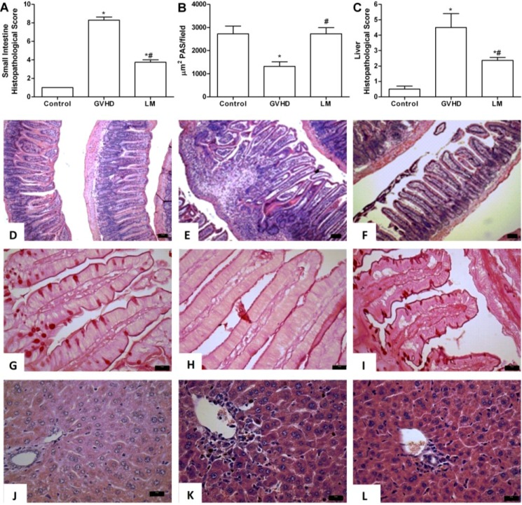
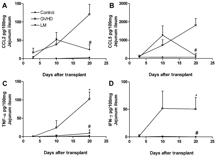
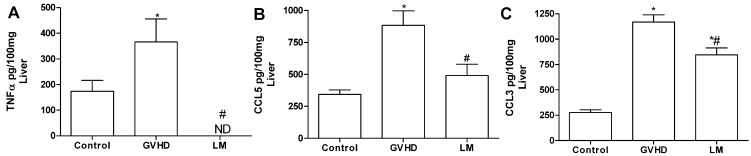
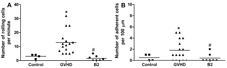
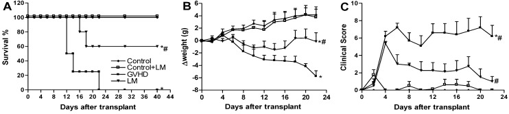
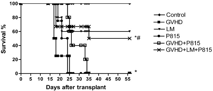
Lithothamnion muelleri (Hapalidiaceae) is a marine red alga, which is a member of a group of algae with anti-inflammatory, antitumor, and immunomodulatory properties. The present study evaluated the effects of treatment with Lithothamnion muelleri extract (LM) in a model of acute graft-versus-host disease (GVHD), using a model of adoptive splenocyte transfer from C57BL/6 donors into B6D2F1 recipient mice. Mice treated with LM showed reduced clinical signs of disease and mortality when compared with untreated mice. LM-treated mice had reduced tissue injury, less bacterial translocation, and decreased levels of proinflammatory cytokines and chemokines (interferon-γ (IFN-γ), tumor necrosis factor-α (TNF-α), chemokine (C-C motif) ligand 2 (CCL2), chemokine (C-C motif) ligand 3 (CCL3) and chemokine (C-C motif) ligand 5 (CCL5)). The polysaccharide-rich fraction derived from LM could inhibit leukocyte rolling and adhesion in intestinal venules, as assessed by intravital microscopy. LM treatment did not impair the beneficial effects of graft-versus-leukaemia (GVL). Altogether, our studies suggest that treatment with Lithothamnion muelleri has a potential therapeutic application in GVHD treatment.


Figures

Figure 1
Figure 1
Lithothamnion muelleri extract (LM) treatment reduces intestinal and hepatic injury in mice subjected to GVHD. GVHD was induced by the adoptive transfer of splenocytes from C57BL/6J donors to B6D2F1 mice. Mice that received syngeneic (B6D2F1) splenocytes did not develop disease and were considered the Control group. LM (1% in the diet, w/w) was offered in the diet of recipient mice from one day before transplantation until the experimental endpoint. At day 20 after transplantation, the mice were sacrificed and the jejunum-ileum and liver tissues were sampled for histopathological analysis (A and C). The numbers of PAS-positive goblet cells counted at 40×/field were determined from four fields per intestine section (B). DF, histological aspects of H&E-stained small intestine sections in Control, GVHD, and LM-treated mice, respectively. Scale bar, 50 μm for all panels. GI, intestine sections with PAS-positive goblet cells from Control, GVHD, and LM treated mice, respectively. JL, histological aspects of H&E-stained liver sections from Control, GVHD, and LM treated mice, respectively. Scale bar, 20 μm for all panels. The results are presented as the mean ± SEM (n = 6); * and #, p < 0.05 when compared with the Control and GVHD groups, respectively.
Figure 2
Figure 2
Lithothamnion muelleri extract (LM) treatment inhibits bacterial translocation and macrophage accumulation in target organs and blood of mice subjected to GVHD. GVHD was induced by the transfer of splenocytes from C57BL/6J donors to B6D2F1 mice. Mice that received syngeneic (B6D2F1) splenocytes did not develop disease and were considered the Control group. LM (1% in the diet, w/w) was offered in the diet of recipient mice from one day before transplantation until the experimental endpoint. At day 20 after transplantation, the mice were sacrificed and the bacterial translocation to the peritoneal cavity (A), blood (B), and liver (C) were quantified. Macrophages were quantified in the jejunum-ileum (D) and liver (E) by enzymatic methods. The results are presented as the mean ± SEM (n = 6); * and #, p < 0.05 when compared with the Control and GVHD groups, respectively.
Figure 3
Figure 3
Lithothamnion muelleri extract (LM) treatment reduces the concentrations of cytokines and chemokines in the small intestine of mice subjected to GVHD. GVHD was induced by the transfer of splenocytes from C57BL/6J donors to B6D2F1 mice. Mice that received syngeneic (B6D2F1) splenocytes did not develop disease and were considered the Control group. LM (1% in the diet, w/w) was offered in the diets of recipient mice from 1 day before transplantation until the experimental endpoint. At 20 days after transplantation, the mice were sacrificed and the concentrations of CCL2 (A), CCL5 (B), TNF-α (C) and IFN-γ (D) in the intestinal homogenates were evaluated by Enzyme Linked Immuno Sorbent Assay (ELISA). The results are shown as the mean ± SEM (n = 5); * and #, p < 0.05 when compared with the Control and GVHD groups, respectively.
Figure 4
Figure 4
Lithothamnion muelleri extract (LM) treatment reduces the concentrations of cytokines and chemokines in the liver of mice subjected to GVHD. GVHD was induced by the transfer of splenocytes from C57BL/6J donors to B6D2F1 mice. Mice that received syngeneic (B6D2F1) splenocytes did not develop disease and were considered the Control group. LM (1% in the diet, w/w) was offered in the diets of recipient mice from one day before transplantation until the experimental endpoint. At 20 days after transplantation, the mice were sacrificed and the concentrations of CCL3 (A), CCL5 (B), and TNF-α (C) in the liver homogenates were evaluated by ELISA. The results are shown as the mean ± SEM (n = 5); * and #, p < 0.05 when compared with the Control and GVHD groups, respectively.
Figure 5
Figure 5
Treatment with a polysaccharide fraction of Lithothamnion muelleri extract (LM) decreases the number of rolling and adherent leukocytes in the mesenteric vasculature of mice subjected to GVHD. GVHD was induced by the transfer of splenocytes from C57BL/6J donors to B6D2F1 mice. Mice that received syngeneic (B6D2F1) splenocytes did not develop disease and were considered the Control group. At day 10 after transplantation, the mice were anesthetized and intestinal venules (±40 μm) were selected in which to count the numbers of rolling and adherent leukocytes by intravital microscopy. Mice that received C57BL/6J splenocytes were treated either with the vehicle or with a polysaccharide fraction of LM (B2, 100 mg/kg) at 30 min before intravital microscopy. (A) Number of rolling cells/minute; (B) Number of adherent cells/100 μm is presented as the mean ± SEM (n = 7–16). Control group (■), GVHD group (▲) and B2 group (●). * and #, p < 0.05 when compared with the Control and GVHD groups, respectively.
Figure 6
Figure 6
Lithothamnion muelleri extract (LM) treatment decreases death, weight loss, and clinical signs in mice subjected to GVHD. GVHD was induced by the transfer of splenocytes from C57BL/6J donors to B6D2F1 mice. Mice that received syngeneic (B6D2F1) splenocytes did not develop disease and were considered the Control group. LM (1% in the diet, w/w) was offered in the diets of recipient mice from one day before transplantation until the experimental endpoint. After the induction of GVHD, the mice were evaluated every two days for survival (A), body weight (B), and clinical scoring (C). The results are shown as the mean ± SEM and the numbers of animals were as follows: Control group (♦), n = 6; Control + LM group (□), n = 6; GVHD group (■), n = 7 and LM group (▼), n = 7. * and #, p < 0.05 when compared with the Control and GVHD groups, respectively.
Figure 7
Figure 7
Lithothamnion muelleri extract (LM) treatment does not interfere with GVL in mice subjected to GVHD. GVHD was induced by the transfer of splenocytes from (C57BL/6J) donors to B6D2F1 mice. GFP+ P815 cells were injected i.v. into B6D2F1 recipients on day 0 of transplantation. Mice that received only syngeneic (B6D2F1) splenocytes did not develop disease and were considered the Control group. The P815 group received only GFP+ P815 cells, and the other groups received splenocytes from (C57BL/6J) donors as well as GFP+ P815 cells. LM(1% in the diet w/w) was offered in the diets of recipient mice from one day before transplantation until the experimental endpoint. After the induction of GVHD, the mice were evaluated every two days for survival. The results are shown as the mean ± SEM. Control group (♦), n=6; GVHD group (■), n=6; LM group (▼), n=6; P815 group (●), n=6; GVHD + P815 group (□), n=7 and GVHD + LM + P815 group (×), n=7. * and #, p < 0.05 when compared with mice that received no tumor cells and mice that received tumor cells but were not subjected to GVHD, respectively.

Similar articles

Cited by

References

    1. Ferrara J.L., Levine J.E., Reddy P., Holler E. Graft-versus-host disease. Lancet. 2009;373:1550–1561. doi: 10.1016/S0140-6736(09)60237-3. - DOI - PMC - PubMed
    1. Pasquini M.C., Wang Z., Horowitz M.M., Gale R.P. 2010 report from the Center for International Blood and Marrow Transplant Research (CIBMTR): Current uses and outcomes of hematopoietic cell transplants for blood and bone marrow disorders. Clin. Transpl. 2010;2010:87–105. - PubMed
    1. Goker H., Hanznedaroglu I.C., Chao N.J. Acute graft-versus-host disease: Pathobiology and management. Exp. Hematol. 2001;29:259–277. doi: 10.1016/S0301-472X(00)00677-9. - DOI - PubMed
    1. Socié G., Blazar B.R. Acute graft-versus-host disease: From the bench to the bedside. Blood. 2009;114:4327–4336. doi: 10.1182/blood-2009-06-204669. - DOI - PMC - PubMed
    1. Westin J.R., Saliba R.M., de Lima M., Alousi A., Hosing C., Qazilbash M.H., Khouri I.F., Shpall E.J., Anderlini P., Rondon G., et al. Steroid-refractory acute GVHD: Predictors and outcomes. Adv. Hematol. 2011;2011:1–8. - PMC - PubMed

Publication types

LinkOut - more resources