Review
. 2013 Jul-Aug;4(4):298-314.
doi: 10.4161/nucl.25751. Epub 2013 Jul 17.

The nuclear envelope LEM-domain protein emerin

Affiliations
Review

The nuclear envelope LEM-domain protein emerin

Jason M Berk et al. Nucleus. 2013 Jul-Aug.

Abstract

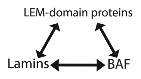
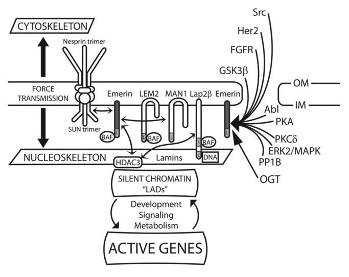
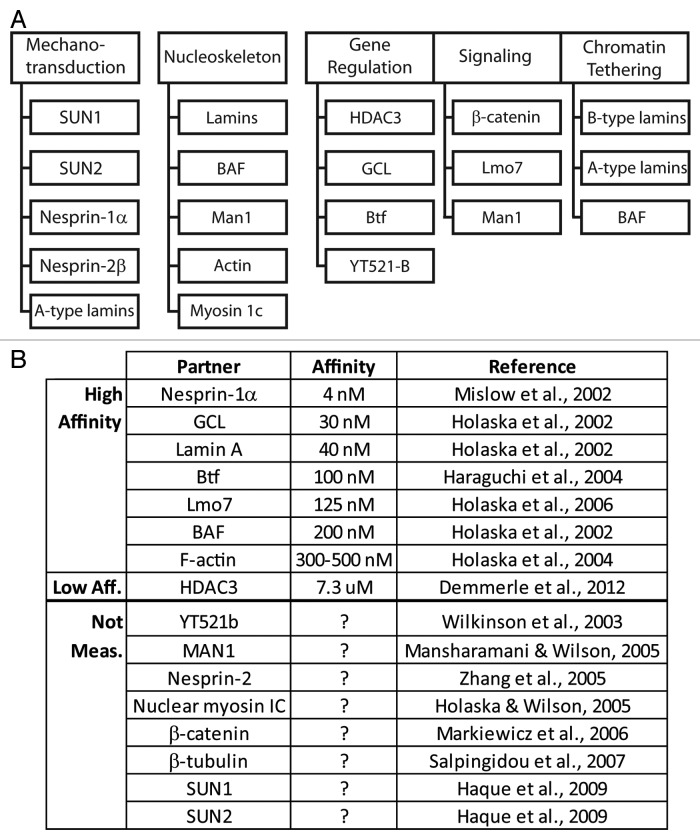
Emerin, a conserved LEM-domain protein, is among the few nuclear membrane proteins for which extensive basic knowledge--biochemistry, partners, functions, localizations, posttranslational regulation, roles in development and links to human disease--is available. This review summarizes emerin and its emerging roles in nuclear "lamina" structure, chromatin tethering, gene regulation, mitosis, nuclear assembly, development, signaling and mechano-transduction. We also highlight many open questions, exploration of which will be critical to understand how this intriguing nuclear membrane protein and its "family" influence the genome.

Keywords: Emery-Dreifuss muscular dystrophy; GAGE cancer-testis antigen; LEM-domain; Nestor-Guillermo progeria; O-GlcNAc transferase; barrier-to-autointegration factor; emerin; laminopathy; nuclear envelope; nucleoskeleton.


Figures

None
Figure 1. Nuclear “lamina” structure has three fundamental components: lamins, LEM-domain proteins and BAF (barrier-to-autointegration factor). These components bind each other with nanomolar affinity in vitro (see text). In C. elegans, loss of any one component (lamin or BAF, or two LEM-domain proteins [Emr-1 and Lem-2]) disrupts co-assembly of the other two and hence blocks nuclear reassembly after mitosis.
None
Figure 2. Schematic depiction of the regulation, partners and selected functions of emerin at the nuclear envelope. Depiction of emerin and other LEM-domain proteins (Lem2, Man1 and Lap2β) at the inner membrane (IM) of the nuclear envelope. Double-headed arrows connect direct binding partners, including emerin (dark gray), SUN-domain proteins, BAF, HDAC3 and Man1. Direct binding to lamins is not indicated. Emerin has roles in signaling, mechano-transduction, nuclear architecture, chromatin tethering and gene regulation. Also depicted are enzymes and pathways that directly target or regulate emerin. “L” indicates the LEM-domain. “L-prime” [L’] in Lap2β indicates the DNA-binding “LEM-like” domain. OGT, O-GlcNAc transferase. OM, outer membrane.
None
Figure 3. Localizations of epitope-tagged emerin polypeptides in cells. Summary of the localization of epitope-tagged emerin polypeptides (truncations, internal deletions or chimeras) expressed transiently in mammalian cells.,WT, wildtype emerin; M, myc-tag; GFP, green fluorescent protein; F, flag tag. Plus or minus indicate polypeptide localization predominately at the nuclear envelope (NE), nucleoplasm (NP) or cytoplasm/endoplasmic reticulum (C/ER), compared with full-length emerin (residues 1–254 or 3–254).
None
Figure 4. Direct binding partners of emerin. (A) Proteins that bind emerin directly in vitro, grouped based on their known or proposed functions in mechanotransduction, nucleoskeleton, gene regulation, signaling or chromatin tethering. (B) Direct partners and equilibrium binding affinity for human emerin in vitro, if known.
None
Figure 5. Functional map based on emerin missense mutations that disrupt binding to specific partners. (A) Summary of binding results for each named partner, tested for binding to each “m-series” (clustered Ala substitutions) or EDMD-causing mutations in emerin (mutations specified in Tables 2 and 3). Scoring: normal binding (+), weakened binding (+/− and gray), and undetectable binding (black box). nt, not tested. (B) Results from (A) mapped schematically to the emerin polypeptide. APC-L, APC-like domain. TM, transmembrane domain. (C) Schematic diagram of known phosphorylation sites in emerin (see Fig. 6). Hexagons, O-GlcNAc sites; circles, phospho-Ser/Thr; squares, phospho-Tyr; white, asynchronous cultures; black, mitotic cultures and conditions. Black with outline, sites identified in both asynchronous and mitotic cells. Double-underlined region has at least two O-GlcNAc sites and potentially other modifications that are uncharacterized due to the large size of the corresponding trypic peptide and poor recovery by mass spectrometry.
None
Figure 6. Published human emerin phosphorylation sites. X indicates emerin phosphorylation sites identified in asynchronous or mitotic cells. Grey columns indicate emerin-specific studies; other columns are high-throughput studies. (S), Ser. (T), Thr. (Y), Tyr. These results are compiled from references –.

References

    1. Dittmer TA, Misteli T. The lamin protein family. Genome Biol. 2011;12:222. doi: 10.1186/gb-2011-12-5-222. - DOI - PMC - PubMed
    1. Simon DN, Wilson KL. The nucleoskeleton as a genome-associated dynamic ‘network of networks’. Nat Rev Mol Cell Biol. 2011;12:695–708. doi: 10.1038/nrm3207. - DOI - PubMed
    1. Schirmer EC, Florens L, Guan T, Yates JR, 3rd, Gerace L. Nuclear membrane proteins with potential disease links found by subtractive proteomics. Science. 2003;301:1380–2. doi: 10.1126/science.1088176. - DOI - PubMed
    1. Wilkie GS, Korfali N, Swanson SK, Malik P, Srsen V, Batrakou DG, et al. Several novel nuclear envelope transmembrane proteins identified in skeletal muscle have cytoskeletal associations. Mol Cell Proteomics. 2011;10:003129. doi: 10.1074/mcp.M110.003129. - DOI - PMC - PubMed
    1. Korfali N, Wilkie GS, Swanson SK, Srsen V, Batrakou DG, Fairley EA, et al. The leukocyte nuclear envelope proteome varies with cell activation and contains novel transmembrane proteins that affect genome architecture. Mol Cell Proteomics. 2010;9:2571–85. doi: 10.1074/mcp.M110.002915. - DOI - PMC - PubMed

Publication types

LinkOut - more resources