. 2013 Jul-Aug;4(4):261-6.
doi: 10.4161/nucl.25341. Epub 2013 Jun 12.

The dynamic nature of the nuclear envelope: lessons from closed mitosis

Affiliations

The dynamic nature of the nuclear envelope: lessons from closed mitosis

James T Arnone et al. Nucleus. 2013 Jul-Aug.

Abstract

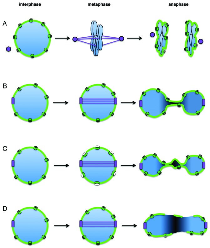
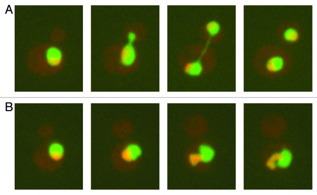
In eukaryotes, chromosomes are encased by a dynamic nuclear envelope. In contrast to metazoans, where the nuclear envelope disassembles during mitosis, many fungi including budding yeast undergo "closed mitosis," where the nuclear envelope remains intact throughout the cell cycle. Consequently, during closed mitosis the nuclear envelope must expand to accommodate chromosome segregation to the two daughter cells. A recent study by Witkin et al. in budding yeast showed that if progression through mitosis is delayed, for example due to checkpoint activation, the nuclear envelope continues to expand despite the block to chromosome segregation. Moreover, this expansion occurs at a specific region of the nuclear envelope- adjacent to the nucleolus- forming an extension referred to as a "flare." These observations raise questions regarding the regulation of nuclear envelope expansion both in budding yeast and in higher eukaryotes, the mechanisms confining mitotic nuclear envelope expansion to a particular region and the possible consequences of failing to regulate nuclear envelope expansion during the cell cycle.

Keywords: checkpoint; mitosis; nuclear envelope; nuclear envelope breakdown; nuclear membrane; nuclear morphology; nucleolus.


Figures

None
Figure 1. The nuclear envelope during different types of mitosis. (A) Open mitosis. During interphase, the chromatin (blue) is contained within the nuclear envelope (light green). As cells enter mitosis, the nuclear envelope disassembles, allowing spindle microtubules (purple lines) nucleated by centrosomes (purple spheres) to align the chromosomes on the metaphase plate. The nuclear envelope reforms in late anaphase, following chromosome segregation. (B) Closed mitosis. Shown is mitosis as it occurs in S. cerevisiae. The spindle pole body (purple) is embedded in the nuclear envelope throughout the cell cycle. After spindle pole body duplication, an intra-nuclear spindle is formed (S. cerevisiae chromosomes do not condense enough to visualize individual chromosomes or a metaphase plate). During anaphase, the nucleus elongates and the nuclear envelope expands as the sister chromatids move away from each other. (C) Functionally open, structurally closed mitosis. A term defined by Sazer to indicate a breakdown in the diffusion barrier while maintaining an intact nuclear envelope. Shown is mitosis as it occurs in Aspergillus nidulans. As in S. cerevisiae, during interphase the spindle pole body is embedded in the nuclear envelope. As cells enter mitosis the nuclear pores (dark green) partially disassemble, significantly reducing the diffusion barrier between the nucleus and cytoplasm. As in closed mitosis, the nucleus elongates to allow chromosome segregation. The bulge in the center of the elongated nucleus during anaphase contains the nucleolus, which is left behind during chromosome segregation. The nuclear pores and the nucleolus reassemble at the end of mitosis. (D) Nuclear envelope rupture. Schizosaccharomyces japonicus cells form an intra-nuclear spindle as in cells undergoing closed or partially open mitosis. However, during anaphase, the nuclear envelope ruptures as the nucleus elongates without nuclear envelope expansion.
None
Figure 2. The budding yeast nucleus throughout the cell cycle. Shown are images taken 20 min apart of budding yeast cells expressing a GFP-tagged nucleoplasmic marker (Pus1) and an RFP-tagged nucleolar marker (Nsr1). (A) Untreated cells. (B) Cells treated with nocodazole. See text for details.

Comment on

  • Witkin KL, Chong Y, Shao S, Webster MT, Lahiri S, Walters AD, Lee B, Koh JL, Prinz WA, Andrews BJ, Cohen-Fix O. The budding yeast nuclear envelope adjacent to the nucleolus serves as a membrane sink during mitotic delay. Curr Biol. 2012;22:1128–33. doi: 10.1016/j.cub.2012.04.022.

References

    1. Hoelz A, Debler EW, Blobel G. The structure of the nuclear pore complex. Annu Rev Biochem. 2011;80:613–43. doi: 10.1146/annurev-biochem-060109-151030. - DOI - PubMed
    1. Scaffidi P, Misteli T. Lamin A-dependent nuclear defects in human aging. Science. 2006;312:1059–63. doi: 10.1126/science.1127168. - DOI - PMC - PubMed
    1. Méndez-López I, Worman HJ. Inner nuclear membrane proteins: impact on human disease. Chromosoma. 2012;121:153–67. doi: 10.1007/s00412-012-0360-2. - DOI - PubMed
    1. Kutay U, Hetzer MW. Reorganization of the nuclear envelope during open mitosis. Curr Opin Cell Biol. 2008;20:669–77. doi: 10.1016/j.ceb.2008.09.010. - DOI - PMC - PubMed
    1. Jaspersen SL, Winey M. The budding yeast spindle pole body: structure, duplication, and function. Annu Rev Cell Dev Biol. 2004;20:1–28. doi: 10.1146/annurev.cellbio.20.022003.114106. - DOI - PubMed

Publication types