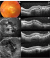
Effect of ranibizumab on serous and vascular pigment epithelial detachments associated with exudative age-related macular degeneration
- PMID: 23874084
- PMCID: PMC3712738
- DOI: 10.2147/DDDT.S46610
Effect of ranibizumab on serous and vascular pigment epithelial detachments associated with exudative age-related macular degeneration
Abstract
Purpose: To report the effect of intravitreal ranibizumab therapy for serous and vascular pigment epithelial detachments (PED) associated with choroidal neovascularisation (CNV) secondary to age-related macular degeneration (AMD).
Methods: In a prospective study, best-corrected visual acuity (BCVA) and optical coherence tomography (OCT) data were collected for 62 eyes of 62 patients, with serous or vascular PED associated with CNV secondary to AMD. Intravitreal ranibizumab 0.5 mg was administered with a loading phase of three consecutive monthly injections, followed by monthly review with further treatment, as indicated according to the retreatment criteria of the PrONTO study. The change in visual acuity and PED height from baseline to month 12 after the first injection was determined.
Results: Sixty-one eyes of 61 patients (one of the patients developed retinal pigment epithelial tear and was excluded from the study) were assessed at the 12-month follow-up examination. There were two types of PED, including vascular PED in 32 patients (Group A) and serous PED (Group B) in 29 patients. The mean improvement of mean BCVA from baseline to 12 months was 0.09 logMAR (Logarithm of the Minimum Angle of Resolution) in Group A and 0.13 logMAR in Group B. Both groups showed significant improvement of the mean BCVA 12 months after the first injection compared with the baseline value (P < 0.05). In relation to the PED height, the mean decrease of mean PED height from baseline to 12 months was 135 μm in Group A and 180 μm in Group B. Both groups showed significant reduction of the PED height during the follow-up period (P < 0.01). The PED anatomical response to ranibizumab was not correlated with the BCVA improvement in any of the groups. Apart from one patient who developed pigment epithelial tear no other complications were documented.
Conclusion: Ranibizumab is an effective and safe treatment for improving vision in patients with serous and vascular PED, although the anatomical response of the PED to ranibizumab may not correlate directly with the visual outcome.
Keywords: age-related macular degeneration; choroidal neovascularisation; intravitreal injection; pigment epithelial detachment; ranibizumab.
Figures


References
-
- Klein R, Klein BE, Jensen SC, Mares-Perlman JA, Cruickshanks KJ, Palta M. Age-related maculopathy in a multiracial United States population: the National Health and Nutrition Examination Survey III. Ophthalmology. 1999;106(6):1056–1065. - PubMed
-
- Klein R, Klein BE, Jensen SC, Meuer SM. The five-year incidence and progression of age-related maculopathy: the Beaver Dam Eye Study. Ophthalmology. 1997;104(1):7–21. - PubMed
-
- Zayit-Soudry S, Moroz I, Loewenstein A. Retinal pigment epithelial detachment. Surv Ophthalmol. 2007;52(3):227–243. - PubMed
-
- Pepple K, Mruthyunjaya P. Retinal pigment epithelial detachments in age-related macular degeneration: classification and therapeutic options. Semin Ophthalmol. 2011;26(3):198–208. - PubMed
-
- Poliner LS, Olk RJ, Burgess D, Gordon ME. Natural history of retinal pigment epithelial detachments in age-related macular degeneration. Ophthalmology. 1986;93(5):543–551. - PubMed
MeSH terms
Substances
LinkOut - more resources
Full Text Sources
Other Literature Sources
Medical

