. 2013;9(7):e1003504.
doi: 10.1371/journal.ppat.1003504. Epub 2013 Jul 18.

Cytotoxic T cells mediate pathology and metastasis in cutaneous leishmaniasis

Affiliations

Cytotoxic T cells mediate pathology and metastasis in cutaneous leishmaniasis

Fernanda O Novais et al. PLoS Pathog. 2013.

Abstract

Disease progression in response to infection can be strongly influenced by both pathogen burden and infection-induced immunopathology. While current therapeutics focus on augmenting protective immune responses, identifying therapeutics that reduce infection-induced immunopathology are clearly warranted. Despite the apparent protective role for murine CD8⁺ T cells following infection with the intracellular parasite Leishmania, CD8⁺ T cells have been paradoxically linked to immunopathological responses in human cutaneous leishmaniasis. Transcriptome analysis of lesions from Leishmania braziliensis patients revealed that genes associated with the cytolytic pathway are highly expressed and CD8⁺ T cells from lesions exhibited a cytolytic phenotype. To determine if CD8⁺ T cells play a causal role in disease, we turned to a murine model. These studies revealed that disease progression and metastasis in L. braziliensis infected mice was independent of parasite burden and was instead directly associated with the presence of CD8⁺ T cells. In mice with severe pathology, we visualized CD8⁺ T cell degranulation and lysis of L. braziliensis infected cells. Finally, in contrast to wild-type CD8⁺ T cells, perforin-deficient cells failed to induce disease. Thus, we show for the first time that cytolytic CD8⁺ T cells mediate immunopathology and drive the development of metastatic lesions in cutaneous leishmaniasis.


Conflict of interest statement

The authors have declared that no competing interests exist.

Figures

Figure 1
Figure 1. Cytolytic profile of CD8+ T cells in lesions from L. braziliensis infected patients.
(A) Heatmap showing induction of key mediators of cytolysis from microarray profiling of ten human lesion biopsies and two normal skin biopsies. Average fold change (FC) for each gene in lesion samples, relative to normal skin controls, is shown, as is the rank for this fold change within the entire expression data set. (B) PBMCs and cells isolated from lesions obtained from L. braziliensis patients were incubated with anti-CD107a. Depicted are representative (A) contour plots and scatter plots of (C) GZMB, (D) PRF and (E) CD107a expression by CD8+ T cells (pregated on Singlets/CD3+/CD8+). Data from patients [8 (blood) and 12 (biopsy)] obtained in two independent experiments are shown. **p<0.01; ***p<0.001.
Figure 2
Figure 2. CD8+ T cells induce pathology during L. braziliensis infection.
(A) BALB/c mice were infected with L. braziliensis in the ear, and the course of infection monitored, and (B) parasite burden assessed in the lesions at 5 weeks. Rag1−/− mice were infected with L. braziliensis in the ear, and reconstituted with either CD8+ T cells, or CD8+ and CD4+ T cells, or did not receive cells and (C) ear thickness at the site of infection assessed. At 7 weeks post infection mice were euthanized and shown are (D) the parasite burden in the lesions and (E) histological comparison of lesions by H&E staining. Scale bar represents 200 µm for lower magnification (top row) and 20 µm for higher magnification (bottom row). (F) Representative front images and back images (showing metastasis) of leishmanial lesions at 6 weeks post infection. Representative data from one of three or more independent experiments (n = 5) with similar results are presented. ns, non-significant. *p<0.05.
Figure 3
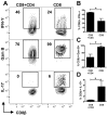
Figure 3. CD8+ T cells from lesions with severe pathology produce IFN-γ, granzyme B and IL-17.
Rag1−/− mice were infected with L. braziliensis in the ear, and reconstituted with either CD8+ T cells, or CD8+ and CD4+ T cells. At 7 weeks post infection mice were euthanized and cells from the lesions were stained for flow cytometry. Depicted are representative (A) contour plots and bar graphs of intracellular (B) IFN-γ, (C) GzmB and (D) IL-17 (pregated on Live/Singlets/CD45+/CD8β+ cells). Bar graphs are representative data from one of three independent experiments (n = 5) with similar results. *p<0.05.
Figure 4
Figure 4. CD8+ T cells degranulate and induce killing of L. braziliensis infected target cells.
(A,B) Rag1−/− mice were infected with L. braziliensis in the ear, and reconstituted with either CD8+ T cells, or CD8+ and CD4+ T cells. Four to six weeks post infection cells isolated from uninfected and infected ears were incubated with anti-CD107a. Depicted are representative (A) contour plots and (B) bar graph of CD107a expression by CD8+ T cells (pregated on Live/Singlets/CD45+/CD8β+ cells). The bar graph represents data from five independent experiments. *p<0.05. (C) Rag1−/− mice were infected with mCherry expressing L. braziliensis in the ear, and reconstituted with eGFP CD8+ T cells. Six weeks post infection cells isolated from infected ears were incubated with anti-CD107a and acquired by ImageStream. Depicted are four representative images showing CD107a expression at the interface between CD8+ T cells and L. braziliensis infected targets. (D) Pie chart with the percentage of eGFP+ CD8+ T cells in contact with infected and uninfected target cells expressing CD107a as assessed by ImageStream. (E) Live-cell imaging of ear cells from Rag1−/− mice infected with mCherry expressing L. braziliensis and reconstituted with eGFP CD8+ T cells four to eight weeks post infection. Numbers represent time in hours∶minutes and scale bar 10 µm. (F) Pie charts with analysis of the outcome of CD8+ T cell and target cell interaction. Data from 63 live-cell imaging fields from 6 independent infection experiments.
Figure 5
Figure 5. CD8+ T cell induced pathology requires specificity.
Rag1−/− mice were infected with L. braziliensis in the ear, and reconstituted with either WT or OT1 CD8+ T cells. (A) Ear thickness was evaluated at the site of infection. (B) Representative images of leishmanial lesions at 6 weeks post infection. At 7 weeks post infection mice were euthanized and shown are: (C) parasite burden in the lesions; (D) contour plots and (E) bar graph of CD107a expression by CD8+ T cells (pregated on: Live/Singlets/CD45+/CD8β+ cells). Graphs are representative data from one of two independent experiments (n = 3) with similar results. *p<0.05.
Figure 6
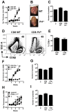
Figure 6. Perforin is required for CD8+ T cell induced pathology in L. braziliensis infection.
Rag1−/− mice were infected with L. braziliensis in the ear, and reconstituted with either WT or Prf−/− CD8+ T cells. (A) Ear thickness at the site of infection. (B) Representative images of leishmanial lesions at 5 weeks post infection. At 7 weeks post infection mice were euthanized and shown are: (C) parasite burden in the lesions; representative (D) contour plots and (E) bar graph of CD8+ T cell in the lesion (pregated on: Live/Singlets/CD45+/CD8β+ cells). Graphs are representative data from one of three independent experiments (n = 5). Rag1−/− mice were infected with L. braziliensis in the ear, and reconstituted with either WT, IFN-γ−/−, or IL-17−/− CD8+ T cells. (F,H) Ear thickness at the site of infection; (G,I) Parasite burden in the lesions at 7 weeks post infection. Data from one (F,G) experiment or one representative experiment of two (H,I) (n = 5) are presented. *p<0.05.

References

    1. Jordan KA, Hunter CA (2010) Regulation of CD8+ T cell responses to infection with parasitic protozoa. Experimental parasitology 126: 318–325. - PMC - PubMed
    1. Gunderson AJ, Mohammed J, Horvath FJ, Podolsky MA, Anderson CR, et al. (2012) CD8(+) T Cells Mediate RAS-Induced Psoriasis-Like Skin Inflammation through IFN-gamma. The Journal of investigative dermatology - PMC - PubMed
    1. Ortega C, Fernandez AS, Carrillo JM, Romero P, Molina IJ, et al. (2009) IL-17-producing CD8+ T lymphocytes from psoriasis skin plaques are cytotoxic effector cells that secrete Th17-related cytokines. Journal of leukocyte biology 86: 435–443. - PubMed
    1. Res PC, Piskin G, de Boer OJ, van der Loos CM, Teeling P, et al. (2010) Overrepresentation of IL-17A and IL-22 producing CD8 T cells in lesional skin suggests their involvement in the pathogenesis of psoriasis. PloS one 5: e14108. - PMC - PubMed
    1. Gebhard JR, Perry CM, Harkins S, Lane T, Mena I, et al. (1998) Coxsackievirus B3-induced myocarditis: perforin exacerbates disease, but plays no detectable role in virus clearance. The American journal of pathology 153: 417–428. - PMC - PubMed

Publication types

MeSH terms