. 2013;9(7):e1003614.
doi: 10.1371/journal.pgen.1003614. Epub 2013 Jul 18.

FGF signalling regulates chromatin organisation during neural differentiation via mechanisms that can be uncoupled from transcription

Affiliations

FGF signalling regulates chromatin organisation during neural differentiation via mechanisms that can be uncoupled from transcription

Nishal S Patel et al. PLoS Genet. 2013.

Erratum in

Abstract

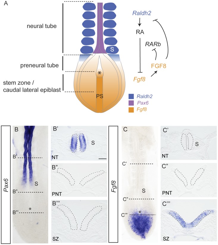
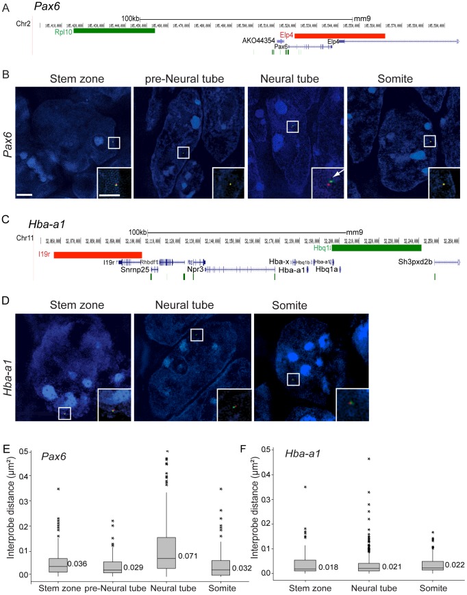
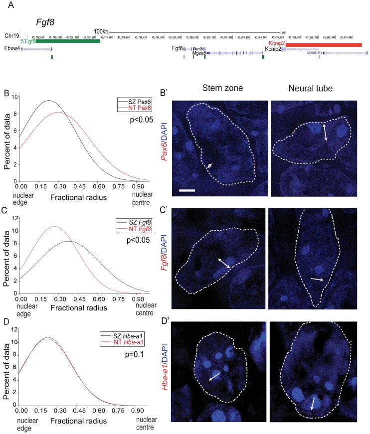
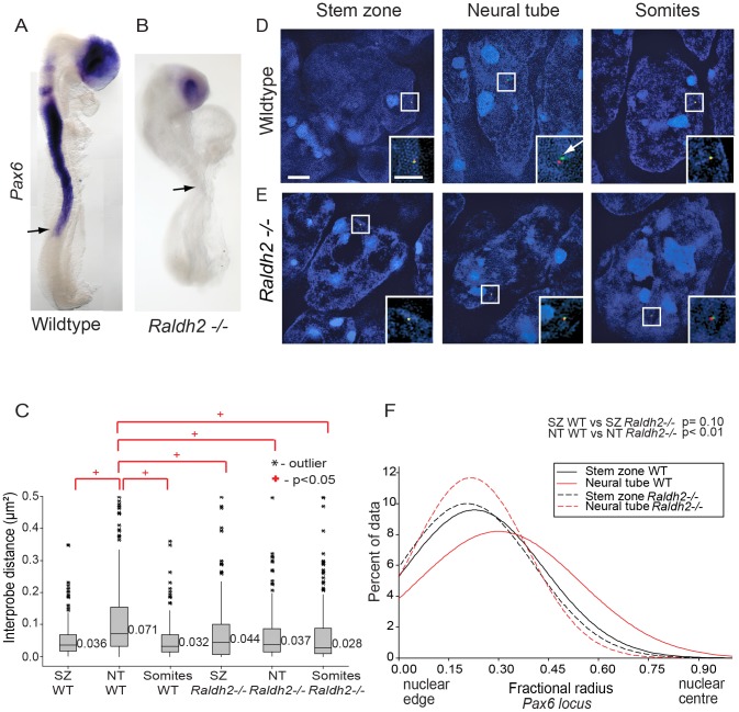
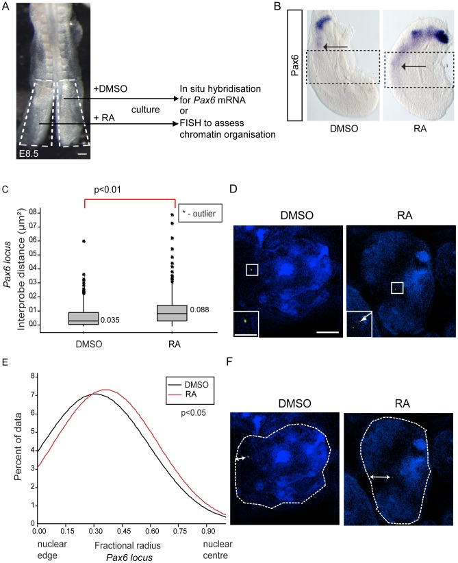
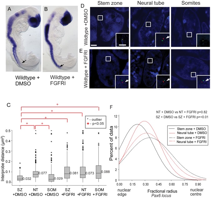
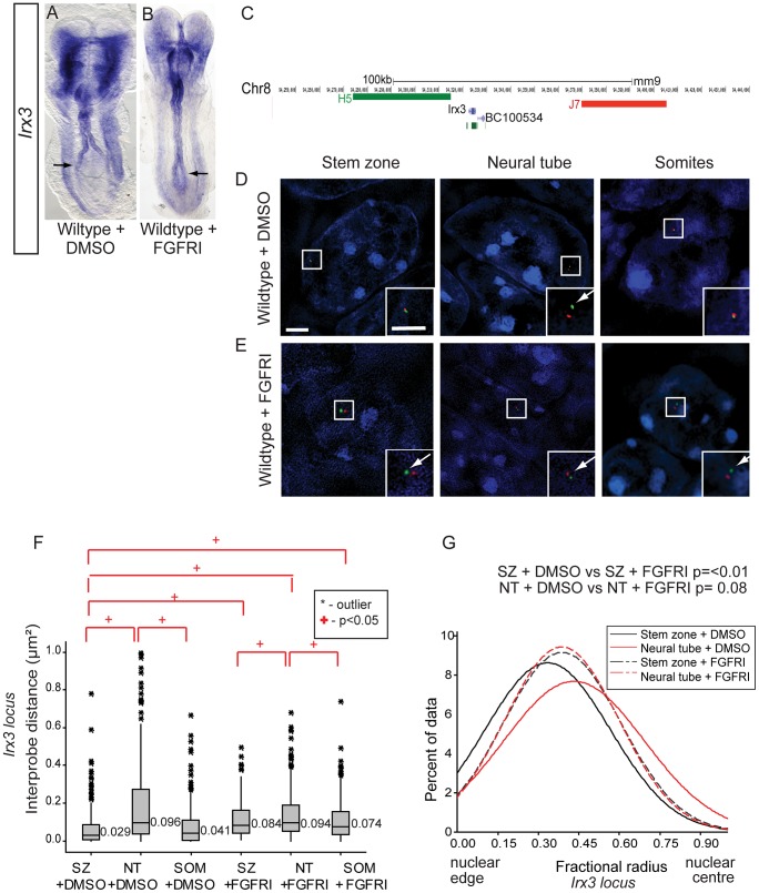
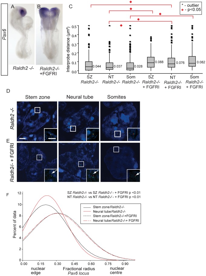
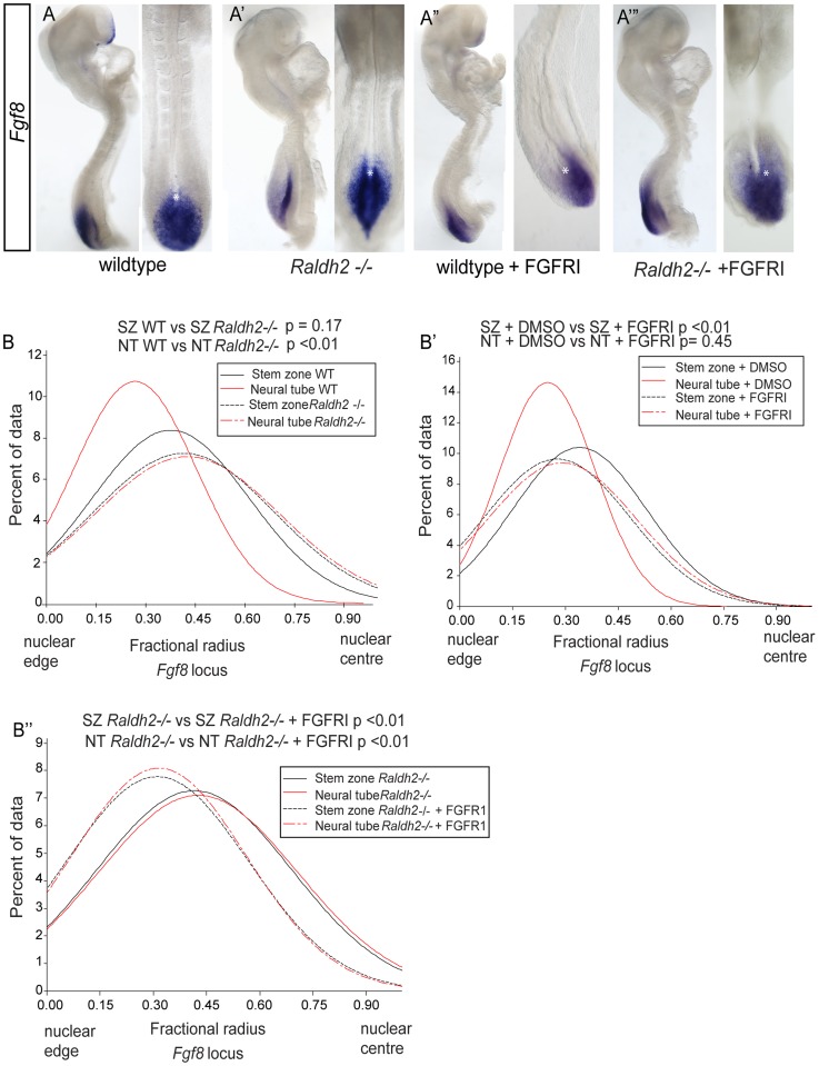
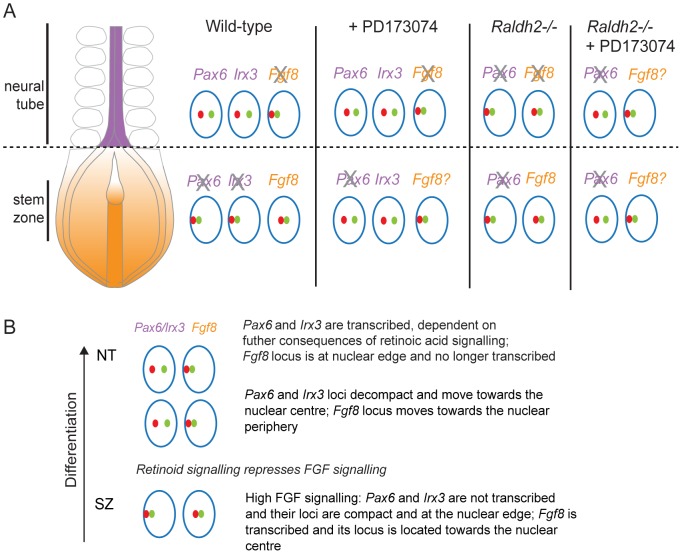
Changes in higher order chromatin organisation have been linked to transcriptional regulation; however, little is known about how such organisation alters during embryonic development or how it is regulated by extrinsic signals. Here we analyse changes in chromatin organisation as neural differentiation progresses, exploiting the clear spatial separation of the temporal events of differentiation along the elongating body axis of the mouse embryo. Combining fluorescence in situ hybridisation with super-resolution structured illumination microscopy, we show that chromatin around key differentiation gene loci Pax6 and Irx3 undergoes both decompaction and displacement towards the nuclear centre coincident with transcriptional onset. Conversely, down-regulation of Fgf8 as neural differentiation commences correlates with a more peripheral nuclear position of this locus. During normal neural differentiation, fibroblast growth factor (FGF) signalling is repressed by retinoic acid, and this vitamin A derivative is further required for transcription of neural genes. We show here that exposure to retinoic acid or inhibition of FGF signalling promotes precocious decompaction and central nuclear positioning of differentiation gene loci. Using the Raldh2 mutant as a model for retinoid deficiency, we further find that such changes in higher order chromatin organisation are dependent on retinoid signalling. In this retinoid deficient condition, FGF signalling persists ectopically in the elongating body, and importantly, we find that inhibiting FGF receptor (FGFR) signalling in Raldh2-/- embryos does not rescue differentiation gene transcription, but does elicit both chromatin decompaction and nuclear position change. These findings demonstrate that regulation of higher order chromatin organisation during differentiation in the embryo can be uncoupled from the machinery that promotes transcription and, for the first time, identify FGF as an extrinsic signal that can direct chromatin compaction and nuclear organisation of gene loci.


Conflict of interest statement

The authors have declared that no competing interests exist.

Figures

Figure 1
Figure 1. Signals regulating differentiation and expression patterns of Pax6 and Fgf8 along the elongating neural axis.
(A) Summary of cell populations, signal localisation and interactions at the caudal end of the E8-8.5 mouse embryo, RA retinoic acid, RAR, retinoic acid receptor, FGF, fibroblast growth factor, PS, primitive streak, S, somite, Raldh2, Retinaldehyde dehydrogenase 2; (B) Pax6 is expressed in the neural tube (B′) in transverse section (TS), but not in (B″) preneural tube or (B‴) stem zone; (C) Fgf8 is expressed in the stem zone, but not in the neural tube (C′) in TS or in (C″) preneural tube, stem zone expression in TS (C‴). Grey dashed lines in B′, B″, B‴, C′,C″, C‴ outline cell populations in which nuclei were assessed in FISH experiments. Scale bar = 50 microns, asterisk indicates position of the node in all embryo images.
Figure 2
Figure 2. Pax6 locus decompaction coincides with Pax6 transcription.
(A) Fosmids (green and red bars) used to analyse chromatin around the Pax6 locus mapped to the mm9 assembly of the mouse genome; (B) Examples of FISH images in DAPI-stained nuclei for the Pax6-flanking probe pairs in stem zone, preneural tube, neural tube, and somite. Insets are enlargements of white boxed areas in each image; (C) Fosmids used to analyse chromatin around the control Hba-a1 locus mapped to mm9; (D) Examples of FISH images in DAPI-stained nuclei for the Hba-a1-flanking probe pairs in stem zone, neural tube, and somite; (E and F) Box-plots of inter-probe distances (µm2) for Pax6 (E) and control Hba-a1 (F) flanking probes in each tissue assessed. Scale bar = 2 microns in examples of FISH images and 1 micron in insets, here and in all subsequent figures.
Figure 3
Figure 3. Pax6 and Fgf8 loci exhibit altered nuclear radial position coincident with transcriptional status.
(A) Fosmids flanking the Fgf8 locus mapped to the mm9 assembly of the mouse genome; (B) Distribution of fractional radius measurements of the Pax6 locus (Elp4 fosmid probe) with respect to the nuclear edge in stem zone (SZ) (black solid line) and neural tube (NT) (red solid line) nuclei. Data are from >50 nuclei per region in each of 3 different embryos; (B′) examples of hybridised nuclei in stem zone and neural tube; (C) Distribution of fractional radius measurements from the Fgf8 locus (Kcnip2 fosmid probe) with respect to the nuclear edge in stem zone and neural tube, showing shift towards nuclear periphery in NT; (C′) examples of hybridised nuclei in stem zone and neural tube; (D) Distribution for fractional radius measurements of control locus Hba-a1 with respect to the nuclear edge in stem zone and neural tube, showing no significant change; (D′) examples of hybridised nuclei in stem zone and neural tube.
Figure 4
Figure 4. Retinoid signalling is required for decompaction around Pax6.
(A) Pax6 is transcribed in wildtype (WT) (B), but not in Raldh2 mutant neural tube (arrows indicate the last formed somite); (C) Box-plots of inter-probe FISH probe distances, in WT and Raldh2−/− embryos showing that in contrast to WT, the Pax6 locus does not decompact in Raldh2 mutant neural tube (NT) and distances remain similar to WT stem zone (SZ) and to somites (S); examples of hybridised nuclei in WT (D) stem zone, neural tube and somites, and in Raldh2 mutant tissues (E) stem zone, neural tube and somites. (F) Graph of data distribution for fractional radius measurements in WT and Raldh2 mutant tissues, showing that the Pax6 locus fails to shift towards the nuclear centre in the neural tube in retinoid deficient conditions.
Figure 5
Figure 5. Exogenous retinoic acid induces Pax6 expression, decompaction and centralised location of Pax6 locus in stem zone explants.
(A) Experimental design, E8.5 embryo bisected along the caudal midline to give an explant pair (white dashed outline), one explant exposed to retinoic acid (RA), the other only to vehicle control (DMSO), followed by analysis for mRNA or chromatin organisation; scale bar = 100 microns (B) Explant pair treated with DMSO or RA analysed for Pax6 expression (caudal limit of expression indicated by black arrows), dotted lines indicate regions examined by FISH for Pax6 inter-probe distance; (C) Inter-probe distances across the Pax6 locus in nuclei taken from sections in the middle third of explants (see Materials and Methods), increased significantly on RA treatment; (D) DAPI stained nuclei and fosmids across the Pax6 locus from explants following exposure to DMSO or RA; (E) Fractional radius measurements (see Materials and Methods) show shift towards nuclear centre following exposure to RA; (F) DAPI stained nuclei and fosmids across the Pax6 locus from explants following exposure to DMSO or RA, showing distance from nuclear edge.
Figure 6
Figure 6. FGF signalling regulates chromatin compaction around Pax6.
(A) Wildtype (WT) embryos exposed to DMSO or (B) FGFR inhibitor (FGFRI) PD173074, showing that blocking FGF elicits Pax6 expression in the preneural tube (arrow = last formed somite); (C) Box-plot of inter-probe distances (µm2) for Pax6 flanking probes in each tissue assessed in DMSO and PD173074 treated embryos, showing that FGFR signalling is required to maintain chromatin compaction around the Pax6 locus in the SZ; examples of hybridised nuclei in DMSO (D) stem zone, neural tube and somites, and PD173074 treated tissues (E) stem zone, neural tube and somites; (F) Graph of data distribution for fractional radius measurements in DMSO and PD173074 treated tissues, showing that the Pax6 locus now shifts towards the nuclear centre in stem zone as well as in the nucleus of neural tube cells after FGFRI treatment.
Figure 7
Figure 7. FGF signalling regulates chromatin compaction and nuclear position at the locus of a further neural progenitor gene, Irx3.
(A) Irx3 is transcribed in the neural tube of wildtype DMSO treated embryos and (B) its expression extends caudally following exposure to PD173074 for 7 h; (C) Fosmids flanking the Irx3 locus mapped to mm9; Examples of FISH images in DAPI-stained nuclei for the Irx3 -flanking probe pairs in stem zone, neural tube, and somite following exposure to (D) DMSO or (E) FGFR inhibitor PD173074; (F) Box-plot of inter-probe distances (µm2) for Irx3 flanking probes in each tissue assessed in DMSO and PD173074 treated embryos, showing that FGFR signalling is required to maintain chromatin compaction around the Irx3 locus in the stem zone; (G) Graph of data distribution for fractional radius measurements in DMSO and PD173074 treated tissues, showing that the Irx3 locus now shifts towards the nuclear centre in stem zone as well as in the nucleus of neural tube cells after FGFRI treatment.
Figure 8
Figure 8. Inhibition of FGFR signalling in Raldh2 mutants rescues higher order chromatin organisation around the Pax6 locus, but not Pax6 transcription.
(A) Pax6 transcripts are lacking in the neural tube of Raldh2 mutants treated with DMSO or (B) with FGFR inhibitor PD173074; (C) Box-plot of inter-probe distances (µm2) for Pax6 flanking probes in each tissue assessed in Raldh2 mutant embryos or Raldh2 mutants treated with PD173074, showing blocking FGFR signalling decompacts the Pax6 locus in both stem zone, neural tube and somites; and examples of hybridised nuclei in Raldh2 mutant embryos, D) stem zone, neural tube and somites, or treated with PD173074, (E) stem zone, neural tube and somites. (F) Graph of data distribution for fractional radius measurements in Raldh2 −/− and Raldh2−/− + PD173074 tissues, showing that the Pax6 locus now shifts towards the nuclear centre in stem zone as well as in the neural tube when FGFR signalling is blocked.
Figure 9
Figure 9. Ectopic Fgf8 expression in Raldh2 mutants correlates with a more central nuclear position, which is dependent on FGF signalling.
Fgf8 expression patterns in (A) wildtype, (A′) Raldh2 −/−, (A″) wildtype treated with FGFR inhibitor, PD173074, (A‴) Raldh2−/− treated with PD173074, asterisk indicates node, note Fgf8 transcripts extend further rostral in Raldh2 −/− embryos; Graphs of data distribution for fractional radius measurements from the Fgf8 locus in (B) WT vs Raldh2 −/− ; (B′) WT DMSO vs FGFRI/PD173074 treated; and (B″) Raldh2 −/− vs Raldh2 −/− + FGFRI/PD173074 treated embryos (for images for Fgf8 fosmids in each condition and tissue, see Figure S5).
Figure 10
Figure 10. Changes in chromatin compaction and nuclear position of Pax6 and Fgf8 loci during neural differentiation and following manipulation of retinoid and/or FGF signalling.
(A) Schematic summarising changes in local chromatin organisation of Pax6, Irx3 and Fgf8 loci as neural differentiation commences in the elongating body axis of wildtype and FGFR signalling deficient (+PD173074) and for Pax6 and Fgf8 in retinoid deficient (Raldh2−/−) mouse embryos and when both these signalling pathways are attenuated. These data indicate that FGF signalling promotes chromatin compaction around Pax6 and Irx3 loci and regulates nuclear position of Pax6, Irx3 and Fgf8 gene loci during neural differentiation. Green and red dots represent flanking fosmid pairs and blue circle the nuclear edge, grey cross indicates likely loss of active Fgf8. (B) Summary of chronological steps towards neural differentiation deduced in this study, from the high FGF signalling context in the stem zone to the onset of neural gene expression in the high retinoid signalling environment of the neural tube.

Similar articles

Cited by

References

    1. Wilson V, Olivera-Martinez I, Storey KG (2009) Stem cells, signals and vertebrate body axis extension. Development 136: 1591–1604. - PubMed
    1. Tzouanacou E, Wegener A, Wymeersch FJ, Wilson V, Nicolas JF (2009) Redefining the progression of lineage segregations during mammalian embryogenesis by clonal analysis. Dev Cell 17: 365–376. - PubMed
    1. Diez del Corral R, Olivera-Martinez I, Goriely A, Gale E, Maden M, et al. (2003) Opposing FGF and retinoid pathways control ventral neural pattern, neuronal differentiation, and segmentation during body axis extension. Neuron 40: 65–79. - PubMed
    1. Naiche LA, Holder N, Lewandoski M (2011) FGF4 and FGF8 comprise the wavefront activity that controls somitogenesis. Proceedings of the National Academy of Sciences of the United States of America 108: 4018–4023. - PMC - PubMed
    1. Vermot J, Gallego Llamas J, Fraulob V, Niederreither K, Chambon P, et al. (2005) Retinoic acid controls the bilateral symmetry of somite formation in the mouse embryo. Science 308: 563–566. - PubMed

Publication types

MeSH terms

LinkOut - more resources