

Exquisite light sensitivity of Drosophila melanogaster cryptochrome
- PMID: 23874218
- PMCID: PMC3715431
- DOI: 10.1371/journal.pgen.1003615
Exquisite light sensitivity of Drosophila melanogaster cryptochrome
Abstract
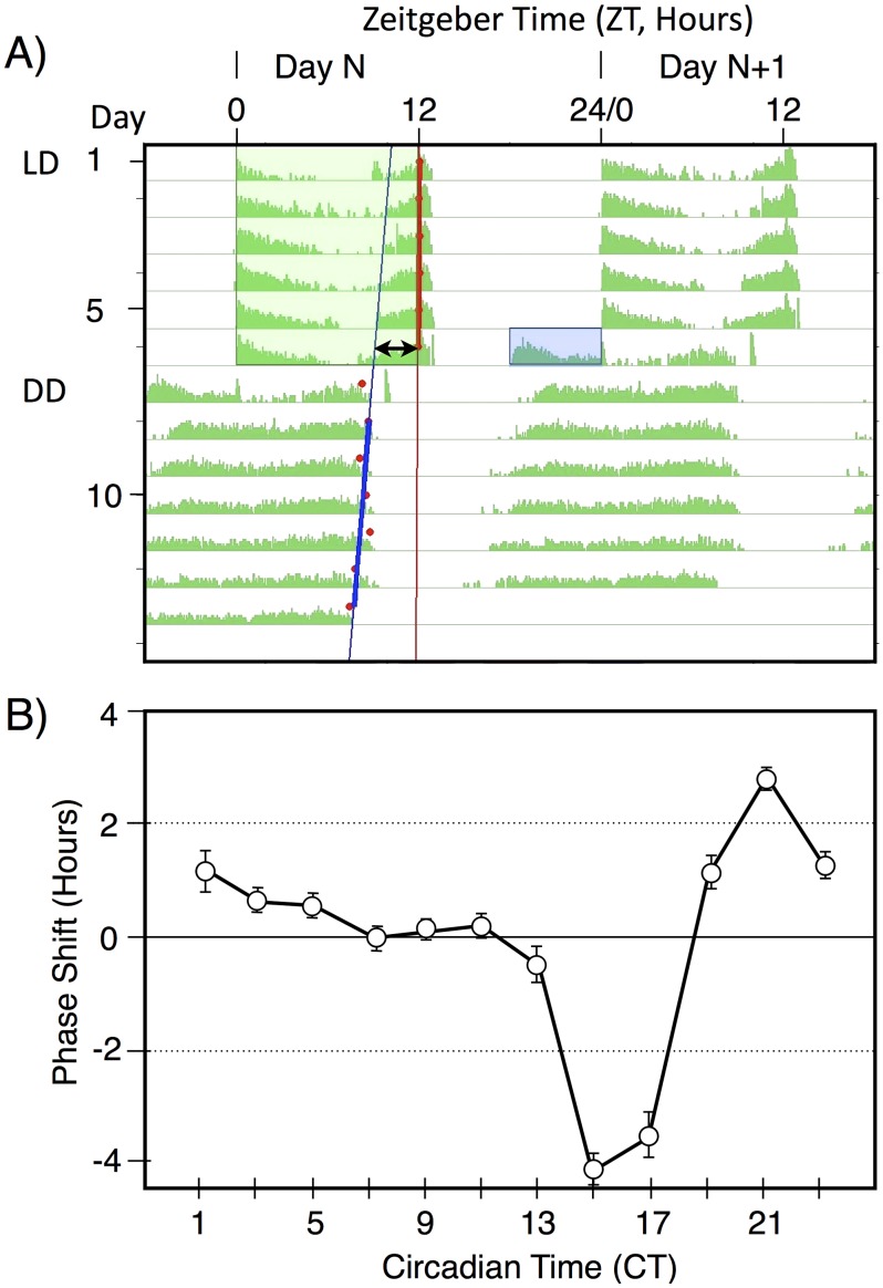
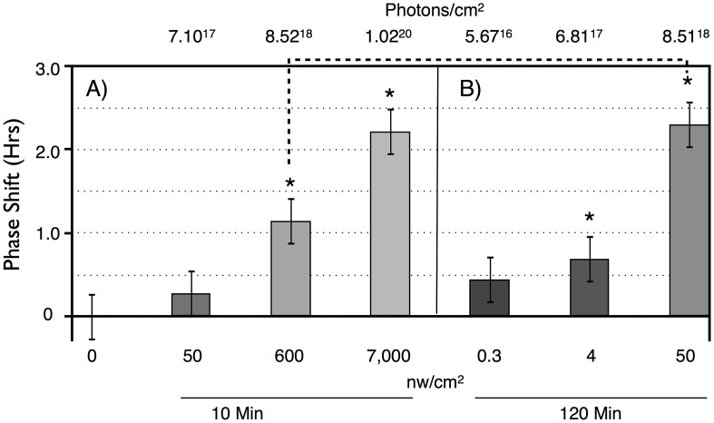
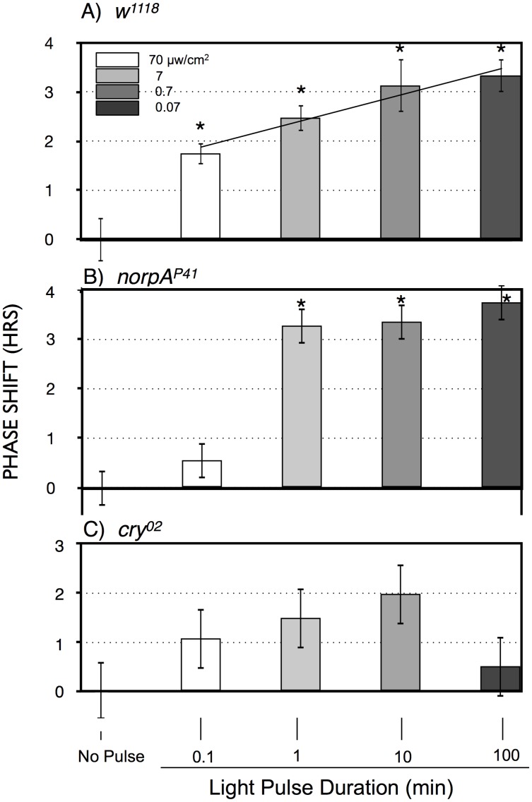
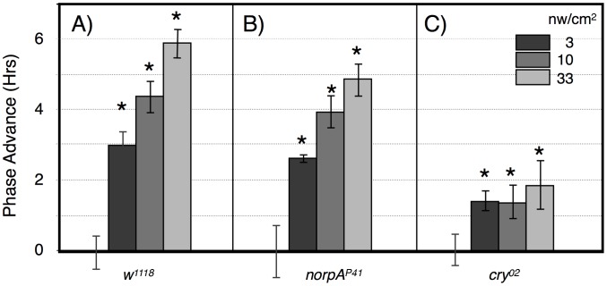
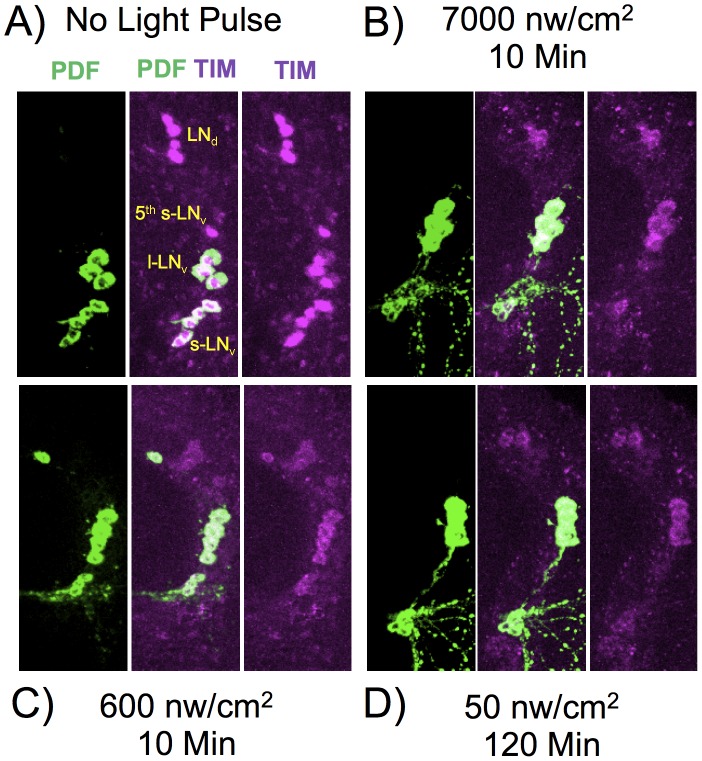
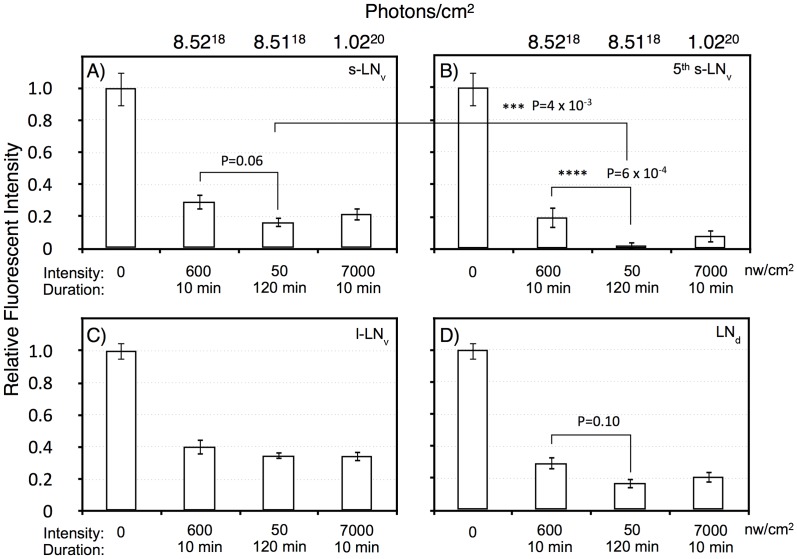
Drosophila melanogaster shows exquisite light sensitivity for modulation of circadian functions in vivo, yet the activities of the Drosophila circadian photopigment cryptochrome (CRY) have only been observed at high light levels. We studied intensity/duration parameters for light pulse induced circadian phase shifts under dim light conditions in vivo. Flies show far greater light sensitivity than previously appreciated, and show a surprising sensitivity increase with pulse duration, implying a process of photic integration active up to at least 6 hours. The CRY target timeless (TIM) shows dim light dependent degradation in circadian pacemaker neurons that parallels phase shift amplitude, indicating that integration occurs at this step, with the strongest effect in a single identified pacemaker neuron. Our findings indicate that CRY compensates for limited light sensitivity in vivo by photon integration over extraordinarily long times, and point to select circadian pacemaker neurons as having important roles.
Conflict of interest statement
The authors have declared that no competing interests exist.
Figures
Similar articles
-
Circadian photoreception in Drosophila: functions of cryptochrome in peripheral and central clocks.J Biol Rhythms. 2001 Jun;16(3):205-15. doi: 10.1177/074873040101600303. J Biol Rhythms. 2001. PMID: 11407780
-
Cryptochrome-positive and -negative clock neurons in Drosophila entrain differentially to light and temperature.J Biol Rhythms. 2010 Dec;25(6):387-98. doi: 10.1177/0748730410381962. J Biol Rhythms. 2010. PMID: 21135155
-
Adult emergence rhythm of fruit flies Drosophila melanogaster under seminatural conditions.J Biol Rhythms. 2012 Aug;27(4):280-6. doi: 10.1177/0748730412448360. J Biol Rhythms. 2012. PMID: 22855572
-
Drosophila Cryptochrome: Variations in Blue.J Biol Rhythms. 2020 Feb;35(1):16-27. doi: 10.1177/0748730419878290. Epub 2019 Oct 10. J Biol Rhythms. 2020. PMID: 31599203 Free PMC article. Review.
-
Blind clocks reveal elusive light input pathway in Drosophila.Trends Neurosci. 2001 Nov;24(11):627-8. doi: 10.1016/s0166-2236(00)01934-2. Trends Neurosci. 2001. PMID: 11672787 Review.
Cited by
-
Cryptochrome-dependent and -independent circadian entrainment circuits in Drosophila.J Neurosci. 2015 Apr 15;35(15):6131-41. doi: 10.1523/JNEUROSCI.0070-15.2015. J Neurosci. 2015. PMID: 25878285 Free PMC article.
-
Circadian light-input pathways in Drosophila.Commun Integr Biol. 2015 Dec 4;9(1):e1102805. doi: 10.1080/19420889.2015.1102805. eCollection 2016 Jan-Feb. Commun Integr Biol. 2015. PMID: 27066180 Free PMC article. Review.
-
Dissecting neuron-specific functions of circadian genes using modified cell-specific CRISPR approaches.Proc Natl Acad Sci U S A. 2023 Jul 18;120(29):e2303779120. doi: 10.1073/pnas.2303779120. Epub 2023 Jul 10. Proc Natl Acad Sci U S A. 2023. PMID: 37428902 Free PMC article.
-
ADHD-associated dopamine transporter, latrophilin and neurofibromin share a dopamine-related locomotor signature in Drosophila.Mol Psychiatry. 2016 Apr;21(4):565-73. doi: 10.1038/mp.2015.55. Epub 2015 May 12. Mol Psychiatry. 2016. PMID: 25962619 Free PMC article.
-
Flies as models for circadian clock adaptation to environmental challenges.Eur J Neurosci. 2020 Jan;51(1):166-181. doi: 10.1111/ejn.14180. Epub 2018 Oct 22. Eur J Neurosci. 2020. PMID: 30269385 Free PMC article. Review.
References
-
- Golombek DA, Rosenstein RE (2010) Physiology of circadian entrainment. Physiol Rev 90: 1063–1102. - PubMed
-
- Peschel N, Helfrich-Forster C (2011) Setting the clock–by nature: circadian rhythm in the fruitfly Drosophila melanogaster. FEBS Lett 585: 1435–1442. - PubMed
-
- Helfrich-Forster C, Winter C, Hofbauer A, Hall JC, Stanewsky R (2001) The circadian clock of fruit flies is blind after elimination of all known photoreceptors. Neuron 30: 249–261. - PubMed
-
- Wheeler DA, Hamblen-Coyle MJ, Dushay MS, Hall JC (1993) Behavior in light-dark cycles of Drosophila mutants that are arrhythmic, blind, or both. J Biol Rhythms 8: 67–94. - PubMed
MeSH terms
Substances
Grants and funding
LinkOut - more resources
Full Text Sources
Other Literature Sources
Molecular Biology Databases
