. 2013;9(7):e1003629.
doi: 10.1371/journal.pgen.1003629. Epub 2013 Jul 11.

Gata4 is required for formation of the genital ridge in mice

Affiliations

Gata4 is required for formation of the genital ridge in mice

Yueh-Chiang Hu et al. PLoS Genet. 2013.

Abstract

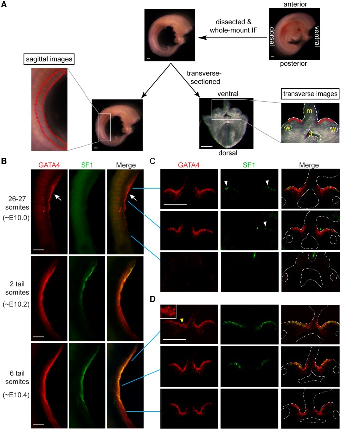
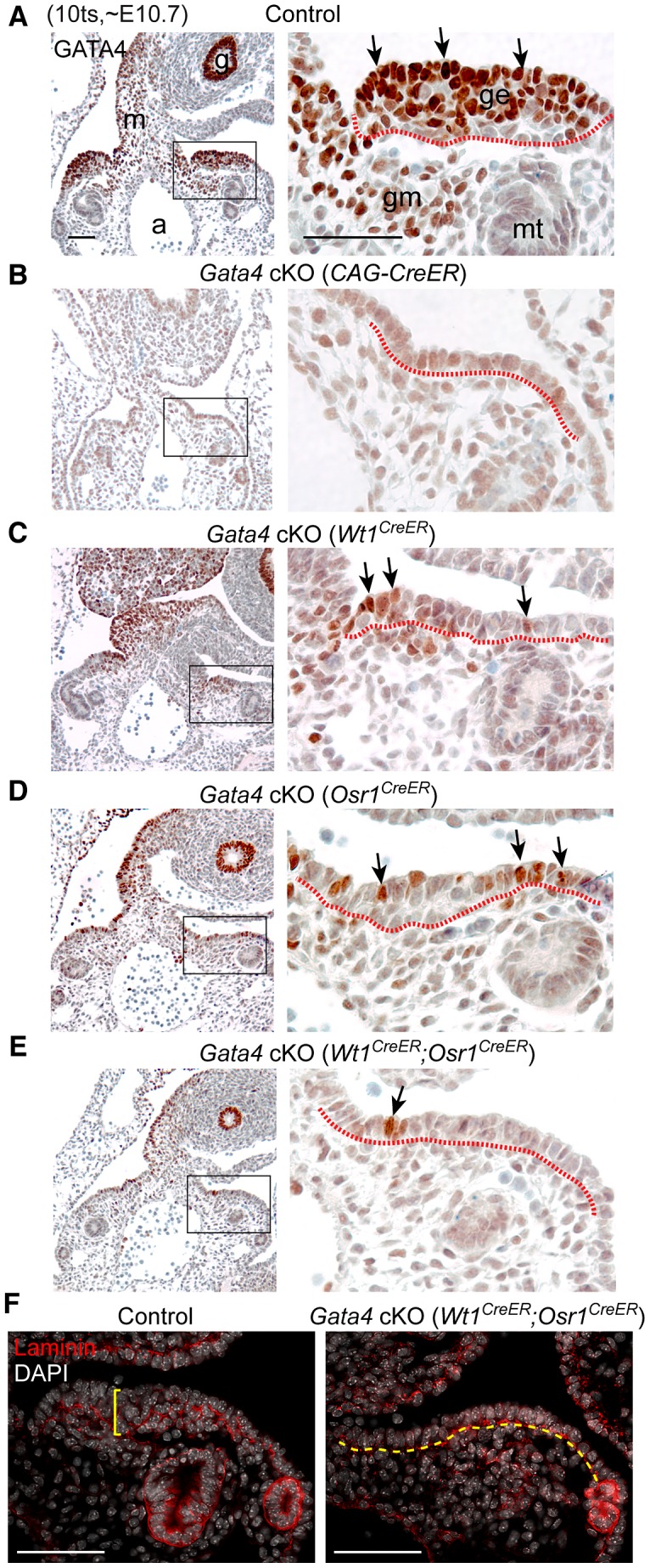
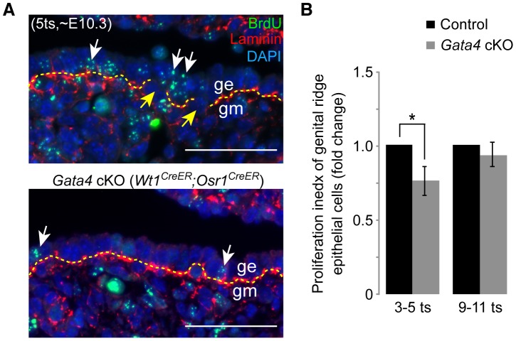
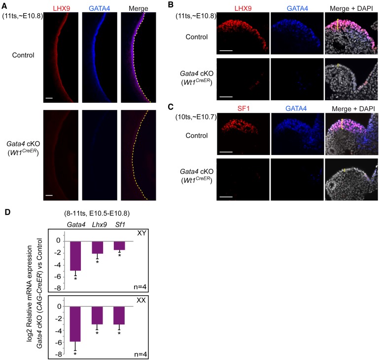
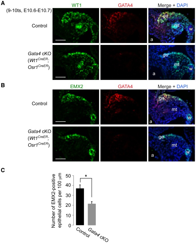
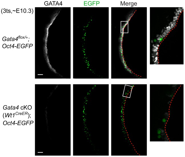
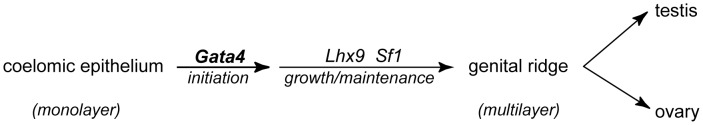
In mammals, both testis and ovary arise from a sexually undifferentiated precursor, the genital ridge, which first appears during mid-gestation as a thickening of the coelomic epithelium on the ventromedial surface of the mesonephros. At least four genes (Lhx9, Sf1, Wt1, and Emx2) have been demonstrated to be required for subsequent growth and maintenance of the genital ridge. However, no gene has been shown to be required for the initial thickening of the coelomic epithelium during genital ridge formation. We report that the transcription factor GATA4 is expressed in the coelomic epithelium of the genital ridge, progressing in an anterior-to-posterior (A-P) direction, immediately preceding an A-P wave of epithelial thickening. Mouse embryos conditionally deficient in Gata4 show no signs of gonadal initiation, as their coelomic epithelium remains a morphologically undifferentiated monolayer. The failure of genital ridge formation in Gata4-deficient embryos is corroborated by the absence of the early gonadal markers LHX9 and SF1. Our data indicate that GATA4 is required to initiate formation of the genital ridge in both XX and XY fetuses, prior to its previously reported role in testicular differentiation of the XY gonad.


Conflict of interest statement

The authors have declared that no competing interests exist.

Figures

Figure 1
Figure 1. GATA4 expression precedes coelomic epithelial thickening and progresses from anterior to posterior.
(A) Schematic representation of experiment. Mouse embryos were dissected to remove limbs, body walls and internal organs, and then subjected to whole-mount immunofluorescence (IF) staining with GATA4 and SF1 antibodies. Stained embryos were imaged sagittally by confocal microscopy, and then transversely (following transverse section), again by confocal microscopy. Red dashed and solid lines in, respectively, sagittal and transverse images indicate location of developing gonads. a, dorsal aorta; m, mesentery; w, Wolffian duct. (B–D) Expression analysis of GATA4 (red) and SF1 (green) protein during early gonadogenesis. GATA4 expression in coelomic epithelia of genital ridges begins in anterior (arrow) and then spreads posteriorly. Epithelial thickening is observed in anterior region of genital ridge at 6-tail-somite stage (yellow arrowhead and inset). SF1 (white arrowheads) is expressed only sporadically in anterior half at 26–27 somite stage. Scale bars: 50 µm.
Figure 2
Figure 2. Gata4 is required for thickening of coelomic epithelium that gives rise to genital ridge.
(A–E) IHC staining for GATA4 protein in transverse sections of control and Gata4 cKO embryos. Sections were chosen to represent similar anterior positions in genital ridges. Right panel shows higher magnification of boxed area in left panel. Arrows indicate examples of positive GATA4 staining. Red dashed lines mark boundary between coelomic epithelium and underlying mesenchyme. Control and conditional mutant embryos, except for Gata4 cKO (CAG-CreER), were from the same litter. a, dorsal aorta; g, gut endoderm; ge, genital ridge epithelium; gm, genital mesenchyme; m, mesentery; mt, mesonephric tubule. (F) Immunofluorescent staining for laminin protein in sections of control and Gata4 cKO embryos. Laminin marks basement membrane. Control genital ridge shows thickened epithelial layer (yellow bracket), whereas the coelomic epithelium in Rb cKO embryo remains a single-cell layer (yellow dashed line). Scale bars: 50 µm.
Figure 3
Figure 3. Gata4 deficiency impairs epithelial proliferation and basement membrane breakdown.
(A) Immunofluorescent staining for BrdU (green) and laminin (red) in sections of control and Gata4 cKO (Wt1CreER;Osr1CreER) embryos where tamoxifen and BrdU were injected at, respectively, E8.75 and ∼E10.0 (6 hours before sacrifice). Yellow dashed lines mark basement membrane. Yellow arrows mark discontinuities in basement membrane in control genital ridge. White arrows indicate representative BrdU-positive epithelial cells. Nuclei stained with DAPI (blue). ge, genital ridge epithelium; gm, genital mesenchyme. Scale bars: 50 µm. (B) Relative proliferation index, comparing the fractions of coelomic epithelial cells positive for BrdU in control and Gata4 cKO (Wt1CreER;Osr1CreER) embryos of the same sex, from the same litter. The index in controls was set at 1. Germ cells were excluded from the counting. At each of the two stages shown, three pairs of control and Gata4 cKO embryos were studied. Plotted here are means ± standard deviation. *, P<0.05 (two-tailed Student's t-test).
Figure 4
Figure 4. Gata4 is required for expression of early gonadal differentiation regulators LHX9 and SF1.
(A–C) Whole-mount immunofluorescent staining for LHX9, SF1, and GATA4 protein in control and Gata4 cKO (Wt1CreER) embryos. Confocal images were taken either sagittally (A) or transversely (B and C). Yellow dashed lines outline coelomic epithelial surface. Yellow brackets indicate thickness of epithelial layer. Scale bars: 50 µm. (D) Quantitative analysis of Gata4, Lhx9, and Sf1 mRNA levels in control and Gata4 cKO (CAG-CreER) urogenital ridges. Plotted here are means ± standard deviation from biological replicates (all values normalized to beta-actin). *, P<0.001 (two-tailed Student's t-test).
Figure 5
Figure 5. Gata4 is not required for expression of WT1 and EMX2 in genital ridge epithelium.
(A and B) Immunofluorescent staining for WT1, EMX2, and GATA4 protein in transverse sections of control and Gata4 cKO (Wt1CreER;Osr1CreER) embryos. a, dorsal aorta; ge, genital ridge epithelium; mt, mesonephric tubule; w, Wolffian duct. Scale bars: 50 µm. (C) Comparison of numbers of EMX2-positive cells per 100 µm (along length of epithelial surface) in control and Gata4 cKO (Wt1CreER;Osr1CreER) embryos. Cells were counted on transverse sections of embryos taken at similar A-P positions. Plotted here are means ± standard deviation from biological replicates (n = 3 for each genotype). *, P = 0.003 (two-tailed Student's t-test).
Figure 6
Figure 6. Migration of primordial germ cells is unaffected in Gata4 cKO embryos.
Whole-mount immunofluorescent staining for GATA4 protein (grey) in control and Gata4 cKO (Wt1CreER) embryos. Confocal images were taken sagittally for a longitudinal view of the genital ridge. Oct4-EGFP transgene marks germ cells (green). Panels on far right provide higher magnification views of boxed areas. Red dashed lines outline the coelomic epithelial surface. Scale bars: 50 µm.
Figure 7
Figure 7. A proposed model for formation of the genital ridge.
Formation of the testis or ovary begins when Gata4-dependent thickening of the coelomic epithelium gives rise to the genital ridge. LHX9 and SF1, acting downstream of GATA4, are subsequently induced, promoting growth and maintenance of the genital ridge. WT1 and EMX2 (not shown), though widely expressed in the genital ridge, are not dependent on Gata4 and likely function in parallel to support growth and maintenance of the genital ridge. The genital ridge then develops as either an ovary or a testis, depending on the sex chromosome constitution of the embryo.

References

    1. Brambell FWR (1927) The development and morphology of the gonads of the mouse - Part I The morphogenesis of the indifferent gonad and of the ovary. Proc R Soc Lond B Biol Sci 101: 391–409.
    1. Gropp A, Ohno S (1966) The presence of a common embryonic blastema for ovarian and testicular parenchymal (follicular, interstitial and tubular) cells in cattle Bos taurus . Z Zellforsch Mikrosk Anat 74: 505–528. - PubMed
    1. Pelliniemi LJ (1975) Ultrastructure of gonadal ridge in male and female pig embryos. Anat Embryol (Berl) 147: 20–34. - PubMed
    1. Wartenberg H, Kinsky I, Viebahn C, Schmolke C (1991) Fine structural characteristics of testicular cord formation in the developing rabbit gonad. J Electron Microsc Tech 19: 133–157. - PubMed
    1. Karl J, Capel B (1998) Sertoli cells of the mouse testis originate from the coelomic epithelium. Dev Biol 203: 323–333. - PubMed

Publication types

Substances