. 2013 Jul 12:5:31.
doi: 10.3389/fnagi.2013.00031. eCollection 2013.

A review of cardiorespiratory fitness-related neuroplasticity in the aging brain

Affiliations

A review of cardiorespiratory fitness-related neuroplasticity in the aging brain

Scott M Hayes et al. Front Aging Neurosci. .

Abstract

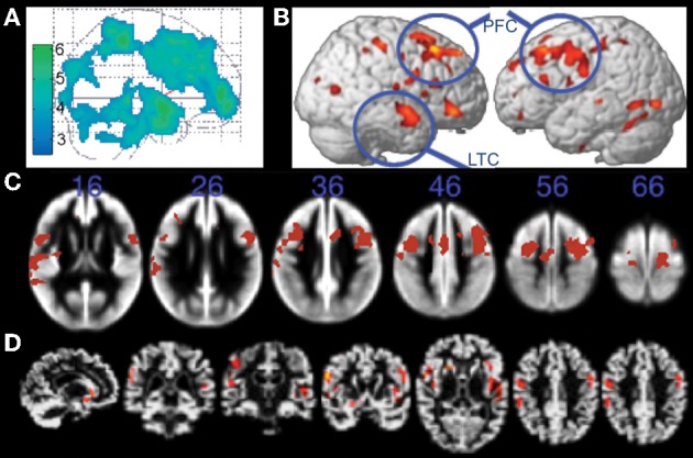
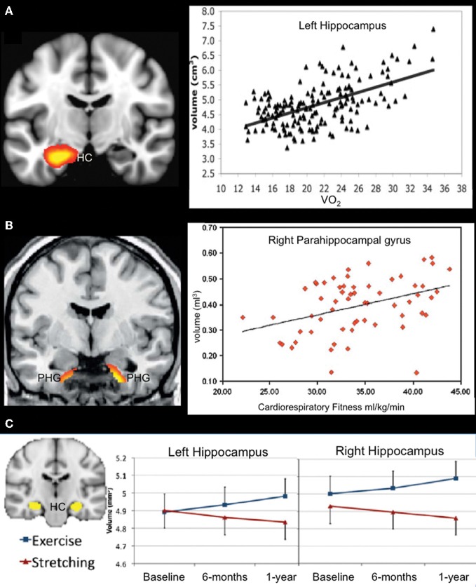
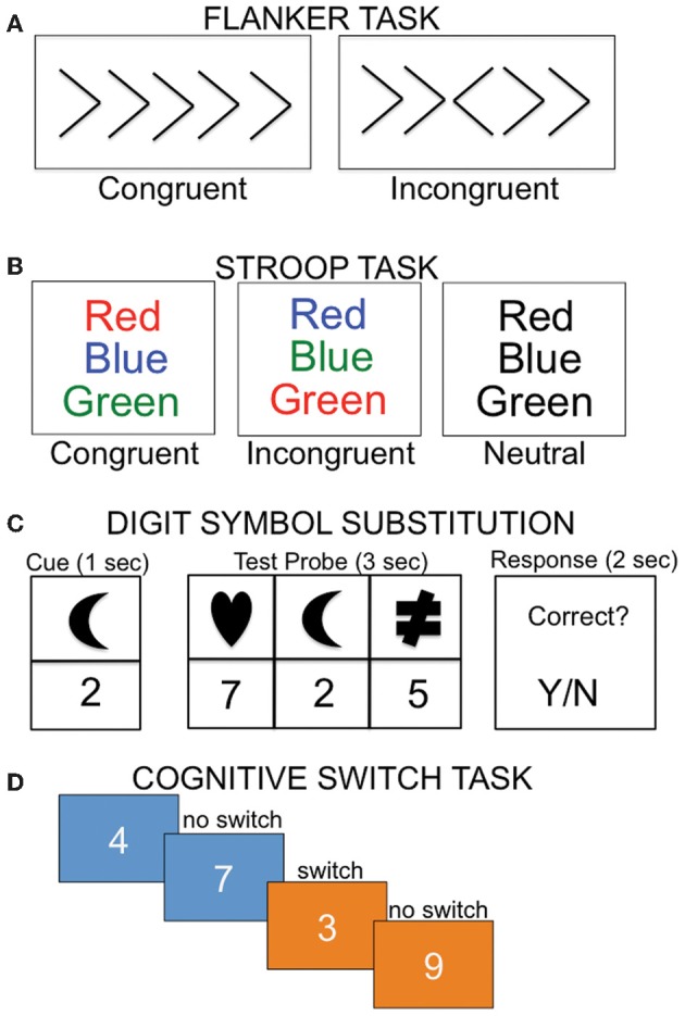
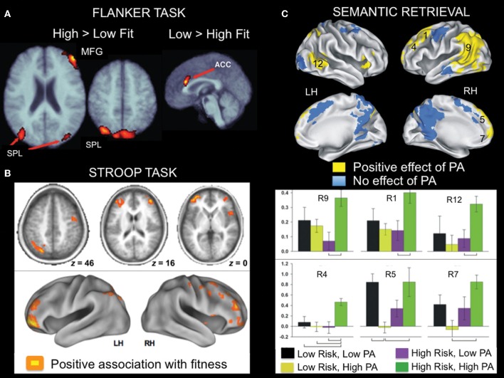
The literature examining the relationship between cardiorespiratory fitness and the brain in older adults has increased rapidly, with 30 of 34 studies published since 2008. Here we review cross-sectional and exercise intervention studies in older adults examining the relationship between cardiorespiratory fitness and brain structure and function, typically assessed using Magnetic Resonance Imaging (MRI). Studies of patients with Alzheimer's disease are discussed when available. The structural MRI studies revealed a consistent positive relationship between cardiorespiratory fitness and brain volume in cortical regions including anterior cingulate, lateral prefrontal, and lateral parietal cortex. Support for a positive relationship between cardiorespiratory fitness and medial temporal lobe volume was less consistent, although evident when a region-of-interest approach was implemented. In fMRI studies, cardiorespiratory fitness in older adults was associated with activation in similar regions as those identified in the structural studies, including anterior cingulate, lateral prefrontal, and lateral parietal cortex, despite heterogeneity among the functional tasks implemented. This comprehensive review highlights the overlap in brain regions showing a positive relationship with cardiorespiratory fitness in both structural and functional imaging modalities. The findings suggest that aerobic exercise and cardiorespiratory fitness contribute to healthy brain aging, although additional studies in Alzheimer's disease are needed.

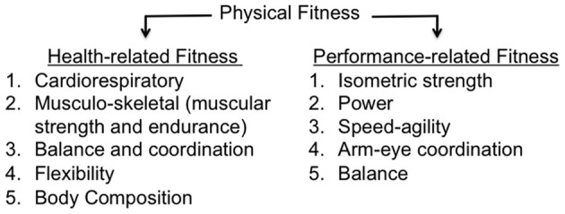
Keywords: diffusion tensor imaging; episodic memory; executive functions; exercise; fMRI; physical activity; physical fitness; structural MRI.


Figures

Figure 1
Figure 1
Taxonomy of physical fitness.
Figure 2
Figure 2
Structural MRI studies showing positive relationships with brain volume and fitness. (A) Gray matter regions, including prefrontal cortex and parietal regions, showing fitness-related preservation in older adults. From Colcombe et al. (2003), Figure 1, p. 178. Adapted with permission. (B) Regions showing increased brain volume in older adults who walked more than 72 blocks per week. From Erickson et al. (2010), Figure 2B, p. 1419. Adapted with permission. (C) Gray matter regions, including bilateral prefrontal cortex, showing a positive relationship with fitness (after controlling for age, gender, and education) in older adults. The blue numbers represent MNI coordinates in the axial (z) plane. From Weinstein et al. (2012), Figure 1A, p. 815. Adapted with permission. (D) Brain regions showing a positive relationship with fitness (after controlling for age, education, and gender) in older adults. From Gordon et al. (2008), Figure 3A, p. 835. Adapted with permission. LTC, lateral temporal cortex; PFC, prefrontal cortex.
Figure 3
Figure 3
Structural MRI studies using a regions-of-interest approach showing a positive relationship between fitness levels and medial temporal lobe volume. (A) Aerobic fitness is associated with bilateral hippocampal volume in older adults (only data from left hippocampus are displayed). From Erickson et al. (2009), Figure 2, p. 1034. Adapted with permission. (B) Increased parahippocampal volume is associated with aerobic fitness in early AD patients. From Honea et al. (2009), Figure 3B, p. 194. Adapted with permission. (C) Aerobic exercise training increases bilateral hippocampal volume in older adults. From Erickson et al. (2011), Figure 1A, p. 3019. Adapted with permission. HC, hippocampus; PHG, parahippocampal gyrus.
Figure 4
Figure 4
Examples of cognitive tasks implemented in imaging studies examining the relationship between cardiorespiratory fitness and the brain. (A) During the flanker task, subjects must indicate the direction of the center arrow, with the incongruent condition requiring additional cognitive control relative to the congruent condition. (B) In the Stroop task, participants are instructed to read the color of the print. In the incongruent condition, participants must inhibit the automatic response of reading the word. (C) In the digit symbol task, participants are required to map the associated number-shape pairs. (D) During the cognitive switching task, subjects are asked to judge whether a number is odd or even when the background is blue, or bigger or smaller than five when the background is orange.
Figure 5
Figure 5
Examples of fMRI studies showing a positive relationship between brain activity and aerobic fitness across a variety of cognitive tasks. (A) Lateral fronto-parietal regions showed increased activation, whereas anterior cingulate cortex showed reduced activation during flanker task performance as a function of fitness in older adults. This pattern was evident in both a cross-sectional and an exercise intervention training study. From Colcombe et al. (2004), Figure 2, p. 3318. Adapted with permission. (B) During Stroop task performance, increased activation in lateral fronto-parietal regions was associated with aerobic fitness. From Prakash et al. (2011), Figure 4, p. 9. Adapted with permission. (C) Brain regions showing greater activation during famous name recognition as a function of physical activity level (PA) in older adults at increased risk for development of AD. The numbers associated with the activation cluster on the surface renderings correspond to the bar graphs presented below. From Smith et al. (2011a), Figures 3, 4, p. 640-641. Adapted with permission. ACC, anterior cingulate cortex; LH, left hemisphere; MFG, middle frontal gyrus; RH, right hemisphere; SPL, superior parietal lobe.

References

    1. Adlard P. A., Perreau V. M., Pop V., Cotman C. W. (2005). Voluntary exercise decreases amyloid load in a transgenic model of Alzheimer's disease. J. Neurosci. 25, 4217–4221 10.1523/JNEUROSCI.0496-05.2005 - DOI - PMC - PubMed
    1. Ashburner J., Friston K. J. (2000). Voxel-based morphometry–the methods. Neuroimage 11, 805–821 10.1006/nimg.2000.0582 - DOI - PubMed
    1. Basser P. J., Pierpaoli C. (1996). Microstructural and physiological features of tissues elucidated by quantitative-diffusion-tensor MRI. J. Magn. Reson. B 111, 209–219 10.1006/jmrb.1996.0086 - DOI - PubMed
    1. Blair S. N., Cheng Y., Holder J. S. (2001). Is physical activity or physical fitness more important in defining health benefits? Med. Sci. Sports. Exerc. 33, S379–S399 discussion S419–S320. - PubMed
    1. Bowles H. R., Fitzgerald S. J., Morrow J. R., Jr., Jackson A. W., Blair S. N. (2004). Construct validity of self-reported historical physical activity. Am. J. Epidemiol. 160, 279–286 10.1093/aje/kwh209 - DOI - PubMed

LinkOut - more resources