. 2013 Jul 12:4:416.
doi: 10.3389/fpsyg.2013.00416. eCollection 2013.

MUC (Memory, Unification, Control) and beyond

Affiliations

MUC (Memory, Unification, Control) and beyond

Peter Hagoort. Front Psychol. .

Abstract

A neurobiological model of language is discussed that overcomes the shortcomings of the classical Wernicke-Lichtheim-Geschwind model. It is based on a subdivision of language processing into three components: Memory, Unification, and Control. The functional components as well as the neurobiological underpinnings of the model are discussed. In addition, the need for extension of the model beyond the classical core regions for language is shown. The attention network and the network for inferential processing are crucial to realize language comprehension beyond single word processing and beyond decoding propositional content. It is shown that this requires the dynamic interaction between multiple brain regions.

Keywords: Control; Memory; Unification; language connectivity; neurobiology of language; speaker meaning.


Figures

Figure 1
Figure 1
The classical Wernicke-Lichtheim-Geschwind model of the neurobiology of language. In this model Broca's area is crucial for language production, Wernicke's area subserves language comprehension, and the necessary information exchange between these areas (such as in reading aloud) is done via the arcuate fasciculus, a major fiber bundle connecting the language areas in temporal cortex (Wernicke's area) and frontal cortex (Broca's area). The language areas are bordering one of the major fissures in the brain, the so-called Sylvian fissure. Collectively, this part of the brain is often referred to as perisylvian cortex.
Figure 2
Figure 2
The MUC model of language. The figure displays a lateral view of the left hemisphere. The numbers indicate Brodmann areas. These are areas with differences in the cytoarchitectonics (i.e., composition of cell types). The memory areas are in the temporal cortex (in yellow) including the angular gyrus in parietal cortex. Unification requires the contribution of Broca's area (Brodmann areas 44 and 45) and adjacent cortex (Brodmann areas 47 and 6) in the frontal lobe. Control operations recruit another part of the frontal lobe (in pink), and the Anterior Cingulate Cortex (ACC; not shown in the figure), as well as areas involved in attention.
Figure 3
Figure 3
The unification gradient in left inferior frontal cortex. Activations and their distribution are shown, related to semantic, syntactic and phonological processing. Regions are based on the meta-analysis in Bookheimer. The centers represent the mean coordinates of the local maxima, the radii represent the standard deviations of the distance between the local maxima and their means. The activation shown is from artificial grammar violations in Petersson et al. (2004) (courtesy of Karl Magnus Petersson).
Figure 4
Figure 4
The arcuate fasciculus in human, chimpanzee and macaque in a schematic lateral view of the left hemisphere. From Rilling et al. (2008); courtesy of Nature Publishing Group.
Figure 5
Figure 5
Simplified illustration of the anatomy and connectivity of the left hemisphere language network. Cortical areas are represented as red circles: pars orbitalis (or), pars triangularis (tr) and pars opercularis (op) of the LIFC; angular gyrus (ag), superior and middle temporal gyri (tg), fusiform gyrus (fg) and temporal pole (tp). White matter fibers are shown in gray, arrows emphasize bi-directional connectivity: arcuate fasciculus (AF), extreme capsule (EC), inferior longitudinal fasciculus (ILF) and uncinate fasciculus (UC). Interfaces with sensory-motor systems are shown in green: visual cortex (vc), auditory cortex (ac) and motor cortex (mc).
Figure 6
Figure 6
The topographical connectivity pattern between frontal and temporal/parietal cortex in the perisylvian language networks. Connections to the left pars opercularis (oper), pars triangularis (tri) and pars orbitalis (orbi) are shown in black, dark gray and white arrows respectively. The solid arrows represent the main (most significant) correlations and the dashed arrows represent the extending (overlapping) connections. Brain areas assumed to be mainly involved in phonological, syntactic and semantic processing are shown in black, dark gray and light gray circles, respectively. P1: Supramarginal gyrus; P3: AG: Angular gyrus; P2: the area between SMG and AG in the superior/inferior parietal lobule; T1: posterior superior temporal gyrus; T2: posterior middle temporal gyrus; P3: inferior temporal gyrus.
Figure 7
Figure 7
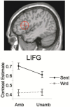
Mean contrast estimated for LIFC for sentences and word sequences, with (Amb) and without (Unamb) noun-verb ambiguities. On top the Region of Interest [ROI; 13 mm sphere around coordinates (−44, 19, 14)] is shown. This ROI includes both BA 44 and parts of BA 45 (Snijders et al., 2009).
Figure 8
Figure 8
Processing cycle subserving word meaning comprehension in the left hemisphere language network. Inputs are conveyed from sensory regions (here visual cortex) to the inferior, middle and superior temporal gyri (1), where lexical information is activated. Signals are hence relayed to the inferior frontal gyrus (2), where neurons respond with a sustained firing pattern. Signals are then fed back into the same regions in temporal cortex from where they were received (3). A recurrent network is thus set up, which allows information to be maintained on-line, a context (green circle) to be formed during subsequent processing cycles, and incoming words to be unified within the context.
Figure 9
Figure 9
Different activations in the four conditions in (A) left superior/inferior parietal cortex; (B) right superior/inferior parietal and right supramarginal region. The gray bars represent the averaged beta values of four conditions in the ROI (the activation in the C–P– condition was taken as an arbitrary zero in the diagram). The vertical lines indicate the standard error for each condition. C+P+: Congruent, with pitch accent; C+P–: Congruent, without pitch accent; C–P+: Incongruent, with pitch accent; C–P–: Incongruent, without pitch accent (from Kristensen et al., 2012).
Figure 10
Figure 10
Illustration of the conditions and the presentation parameters of the fMRI stuy on indirect requests (IR). The top half shows the time course of presentation. On each trial a fixation cross was presented for 500 ms, followed by a visual scene. The utterance was presented auditorily, 200 ms after picture onset. Each trial lasted 3 sec. The bottom half depicts one item in the four conditions. For further details, see (van Ackeren et al., 2012).
Figure 11
Figure 11
Regions of interest were interrogated with respect to the conditions IR, PC, UC, and BC. The image shows all ROI's, superimposed on a brain template. The bar diagrams illustrate mean percent signal change for each condition. The error bars depict the standard error. (A) Green ROIs show regions from the ToM localizer (mPFC and TPJ). (B) Red ROIs refer to regions that were activated during action execution (pre-SMA and bilateral IPL) (van Ackeren et al., 2012).

Similar articles

Cited by

References

    1. Anwander A., Tittgemeyer M., von Cramon D. Y., Friederici A. D., Knosche T. R. (2007). Connectivity-based parcellation of Broca's area. Cereb. Cortex 17, 816–825 10.1093/cercor/bhk034 - DOI - PubMed
    1. Baggio G., Hagoort P. (2011). The balance between memory and unification in semantics: a dynamic account of the N400. Lang. Cogn. Process. 26, 1338–1367 10.1080/01690965.2010.542671 - DOI
    1. Baggio G., Van Lambalgen M., Hagoort P. (2008). Computing and recomputing discourse models: an ERP study. J. Mem. Lang. 59, 36–53 10.1016/j.jml.2008.02.005 - DOI
    1. Bašnáková J., Weber K., Petersson K. M., van Berkum J., Hagoort P. (2013). Beyond the language given: the neural correlates of inferring speaker meaning. Cereb. Cortex. [Epub ahead of print]. 10.1093/cercor/bht112 - DOI - PubMed
    1. Binder J. R., Desai R. H. (2011). The neurobiology of semantic memory. Trends Cogn. Sci. 15, 527–536 10.1016/j.tics.2011.10.001 - DOI - PMC - PubMed

LinkOut - more resources