. 2013 Jul 9;8(7):e67873.
doi: 10.1371/journal.pone.0067873. Print 2013.

Dendritic cell immunoreceptor is a new target for anti-AIDS drug development: identification of DCIR/HIV-1 inhibitors

Affiliations

Dendritic cell immunoreceptor is a new target for anti-AIDS drug development: identification of DCIR/HIV-1 inhibitors

Alexandra A Lambert et al. PLoS One. .

Abstract

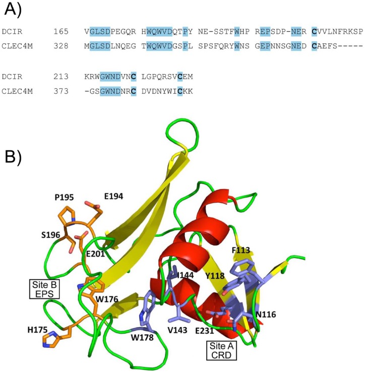
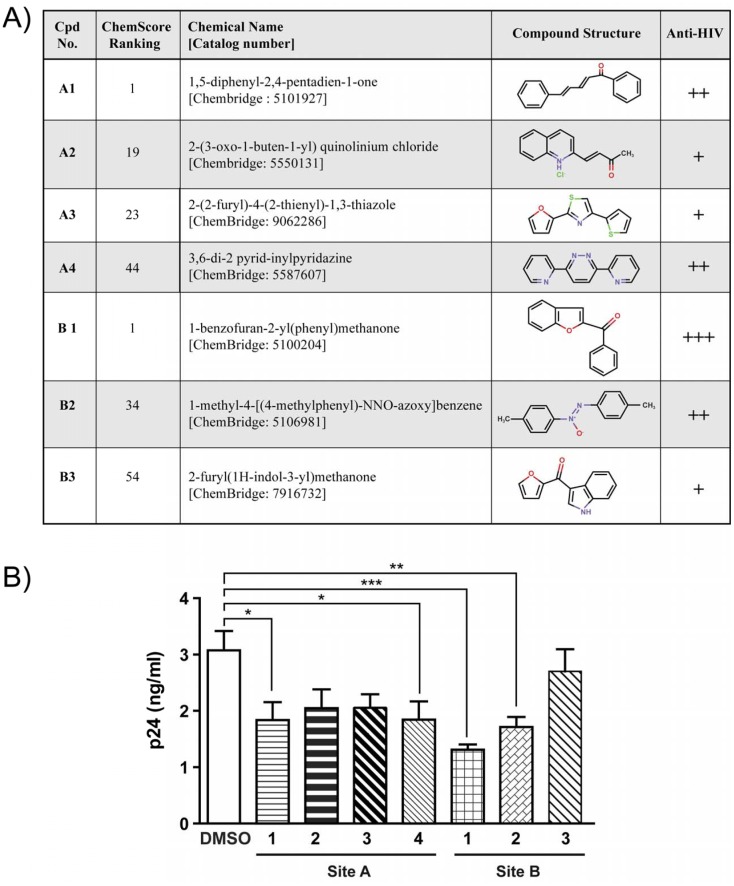
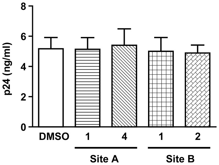
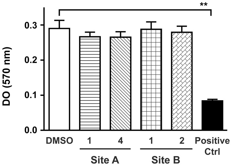
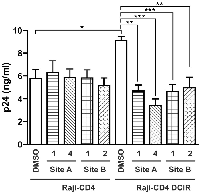
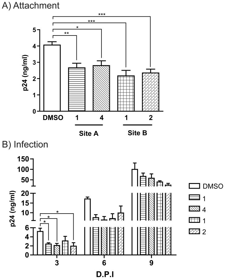
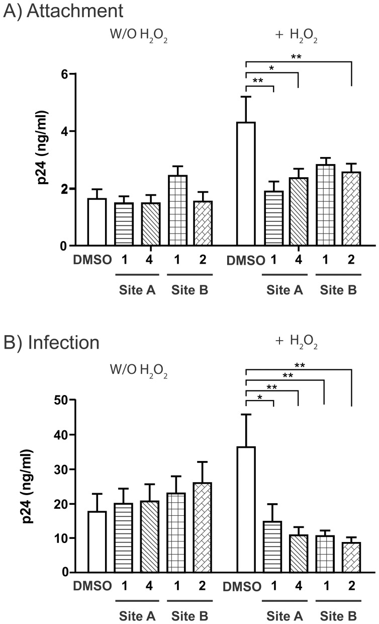
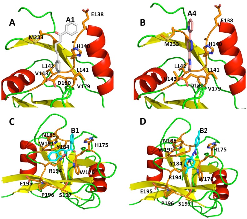
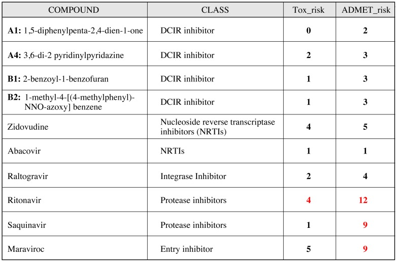
The HIV-1 pandemic continues to expand while no effective vaccine or cure is yet available. Existing therapies have managed to limit mortality and control viral proliferation, but are associated with side effects, do not cure the disease and are subject to development of resistance. Finding new therapeutic targets and drugs is therefore crucial. We have previously shown that the dendritic cell immunoreceptor (DCIR), a C-type lectin receptor expressed on dendritic cells (DCs), acts as an attachment factor for HIV-1 to DCs and contributes to HIV-1 transmission to CD4(+) T lymphocytes (CD4TL). Directly involved in HIV-1 infection, DCIR is expressed in apoptotic or infected CD4TL and promotes trans-infection to bystander cells. Here we report the 3D modelling of the extracellular domain of DCIR. Based on this structure, two surface accessible pockets containing the carbohydrate recognition domain and the EPS binding motif, respectively, were targeted for screening of chemicals that will disrupt normal interaction with HIV-1 particle. Preliminary screening using Raji-CD4-DCIR cells allowed identification of two inhibitors that decreased HIV-1 attachment and propagation. The impact of these inhibitors on infection of DCs and CD4TL was evaluated as well. The results of this study thus identify novel molecules capable of blocking HIV-1 transmission by DCs and CD4TL.


Conflict of interest statement

Competing Interests: The authors have declared that no competing interests exist.

Figures

Figure 1
Figure 1. DCIR modelling.
A) Partial sequence alignment between CLEC4M (GenBank™ #AAI10615) and DCIR (GenBank™ # NP_057268) indicating completely conserved regions and residues. B) Three-dimensional model showing positions of the selected docking sites on the dendritic cell immunoreceptor (DCIR) model (103-233) for virtual screening runs according to the examples. Residues forming site A are represented by the blue portions while orange indicates those forming site B.
Figure 2
Figure 2. Selected compounds screened and tested in vitro.
A) Chemical structure of potential inhibitors of HIV-1 attachment to DCIR, selected by virtual screening, Compounds selected from CRD site are depicted as A1 to A4. Those selected from EPS site are named as B1 to B3 Virtual interacting compounds with both site of DCIR modeling have anti-HIV activity. B) Inhibitors decrease HIV-1 attachment to DCIR: Raji-CD4-DCIR cells were treated with one of four site-A inhibitors or one of three site-B inhibitors (inhibitor concentration = 10 µM) or with solvent (10 mM DMSO) only for 10 min at 37°C. Cells were thereafter exposed to NL4-3 virus for 60 min. After three washes with PBS to remove un-adsorbed virus, the abundance of cell-associated virions was quantified by measuring p24 content. Data correspond to mean ± SEM of three independent experiments performed with triplicate samples. Asterisks (*) denote statistically significant values (*, P<0.05, ** P<0.01, *** P<0.001).
Figure 3
Figure 3. DCIR inhibitors do not affect other C-type lectin binding such as DC-SIGN.
Raji-DC-SIGN cells were treated for 10 min at 37°C with inhibitors 1 and 4 directed against site A and 1 and 2 directed against site B, or with DMSO. Afterwards, cells were pulsed with NL4-3 for 60 min, rinsed thrice with PBS to remove un-adsorbed virus, and cell-associated viruses were quantified by measuring p24 content. Data shown correspond to the means ± SEM of 3 independent experiments performed in triplicate.
Figure 4
Figure 4. Impact of inhibitors on lymphocytes proliferation.
PBMC (2×10⁵ cells/200 µl) were pre-incubated with DCIR inhibitors (10 µM) (or vehicle), or with a toxic molecule as a positive control at 100 uM (Ctrl) before mitogenic stimulation with PHA-L/IL-2 (1 µg/ml and 30 U/ml). Cell proliferation was stopped at day 3 by adding MTT reagent and SDS as described in “Materials and Methods”. A 570 was then determined. Data shown correspond to the means ± SEM of 3 independent experiments performed in triplicate. Asterisks denote statistically significant values (**, P<0.01).
Figure 5
Figure 5. HIV-1 replication is diminished in DCIR-expressing cells by inhibitors specific for the site A and site B.
Raji-CD4 and Raji-CD4-DCIR were treated with the inhibitors selected in figure 2, or DMSO. Cells were then exposed to NL4-3 for 2 h, rinsed and maintained in culture for 3d. Cell-free culture supernatants were collected and assayed for p24 content. Data shown correspond to the means ± SEM from 3 independent experiments performed with triplicate samples. Asterisks denote statistically significant values (*, P<0.05, ** P<0.01, *** P<0.001).
Figure 6
Figure 6. DCIR inhibitors reduce HIV-1 attachment and infection on primary dendritic cells.
A) IM-MDDCs were treated with four chemical inhibitors or DMSO for 10 min at 37°C. Next, cells were pulsed with NL4-3balenv for 60 min at 37°C and rinsed thoroughly before measuring p24 content. B) In some experiments, similarly treated IM-MDDCs were pulsed with NL4-3balenv for 2 h at 37°C, rinsed thoroughly, and maintained in complete culture medium supplemented with GM-CSF and IL-4 for up to 9 days with medium replenishment every 3 days. Cell-free culture supernatants were quantified by measuring p24 content. Data shown correspond to the means ± SEM of 3 independent experiments performed in triplicate. Asterisks denote statistically significant values (*, P<0.05; **, P<0.01; ***, P<0.001).
Figure 7
Figure 7. Impact of DCIR inhibitors on HIV-1 transmission by apoptotic CD4+ T cells.
Target CD4+ T cells (1×10⁶) were treated for 16 h with H2O2 (30 µM) to induce apoptosis and the surface expression of DCIR. Cells (± H2O2 treatment) were incubated for 10 min with a site A inhibitor (1 and 4) or a site B inhibitor (1 and 2) or with 10 mM DMSO. A) Cells were next exposed to NL4-3 for 1 h at 37°C, washed thoroughly to remove un-adsorbed virions before assessing p24 content. B) Cells were first incubated with NL4-3 for 2 h at 37°C, washed thoroughly to remove un-adsorbed virions and cultured with autologous PHA-L/IL-2-activated CD4TL in complete RPMI-1640 supplemented with rhIL-2. Cell-free supernatants were collected on day 3 and assayed for p24 content. Data correspond to mean ± SEM of 4 independent experiments performed in triplicate for panel A and mean ± SEM of 4 independent experiments for panel B. Asterisks denote statistically significant values (*, P<0.05; **, P<0.01).
Figure 8
Figure 8. Three-dimensional schematic representation of the docking pose of selected active molecules.
Panel A: Compound A1 (1,5-diphenyl-2,4-pentadien-1-one) in site A. Panel B: Compound A4∶3,6-di(2 pyridyl) pyridazine in site A. Panel C: Compound B1 (1-benzofuran-2-yl-phenylmethanone in site B. Panel D: Compound B2 (1-methyl-4-[(4-methylphenyl)-NNO-azoxy]benzene) in site B.
Figure 9
Figure 9. ADMET.
Modelled ADMET (Absorption, Distribution, Metabolism, Elimination and Toxicity) properties of our first four lead compounds using ADMET predictor. These results shown that toxicity of our molecules is minimal compare to protease inhibitor or Maraviroc, both molecules currently used in clinic.

References

    1. Deeks SG, Phillips AN (2009) HIV infection, antiretroviral treatment, ageing, and non-AIDS related morbidity. BMJ 338: a3172. - PubMed
    1. Evans DT, Bricker JE, Sanford HB, Lang S, Carville A, et al. (2005) Immunization of macaques with single-cycle simian immunodeficiency virus (SIV) stimulates diverse virus-specific immune responses and reduces viral loads after challenge with SIVmac239. J Virol 79: 7707–7720. - PMC - PubMed
    1. Haase AT (2010) Targeting early infection to prevent HIV-1 mucosal transmission. Nature 464: 217–223. - PubMed
    1. Blanchet F, Moris A, Mitchell JP, Piguet V (2011) A look at HIV journey: from dendritic cells to infection spread in CD4+ T cells. Curr Opin HIV AIDS 6: 391–397. - PubMed
    1. Blanchet FP, Moris A, Nikolic DS, Lehmann M, Cardinaud S, et al. (2010) Human immunodeficiency virus-1 inhibition of immunoamphisomes in dendritic cells impairs early innate and adaptive immune responses. Immunity 32: 654–669. - PMC - PubMed

Publication types

MeSH terms