. 2013 Jul 9;8(7):e67990.
doi: 10.1371/journal.pone.0067990. Print 2013.

β5 integrin up-regulation in brain-derived neurotrophic factor promotes cell motility in human chondrosarcoma

Affiliations

β5 integrin up-regulation in brain-derived neurotrophic factor promotes cell motility in human chondrosarcoma

Chih-Yang Lin et al. PLoS One. .

Abstract

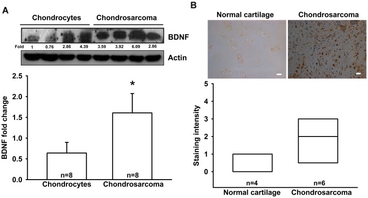
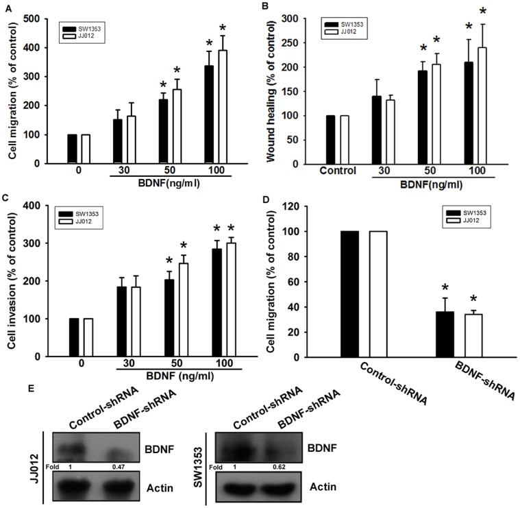
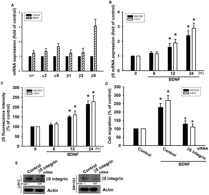
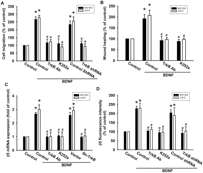
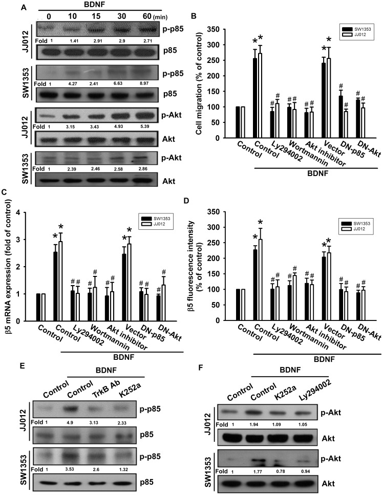
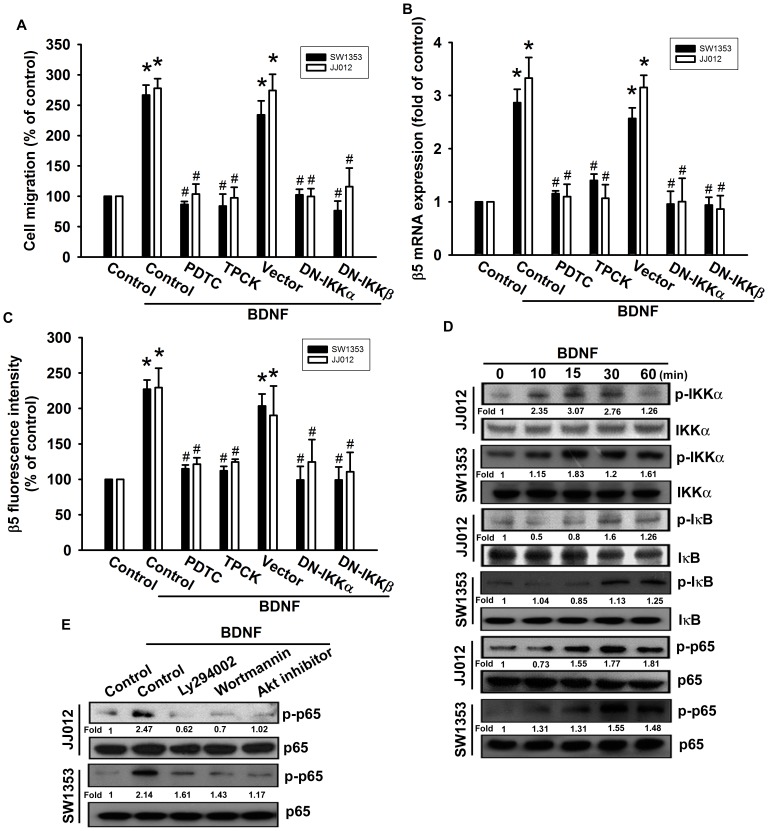
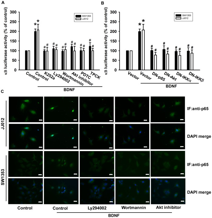
Chondrosarcoma is a primary malignant bone cancer, with a potent capacity to invade locally and cause distant metastasis; it has a poor prognosis and shows a predilection for metastasis to the lungs. Brain derived neurotrophic factor (BDNF) is a small-molecule protein from the neurotrophin family of growth factors that is associated with the disease status and outcomes of cancers. However, the effect of BDNF on migration activity in human chondrosarcoma cells is mostly unknown. Here, we found that human chondrosarcoma tissues showed significant expression of BDNF, which was higher than that in normal cartilage and primary chondrocytes. We also found that BDNF increased the migration and expression of β5 integrin in human chondrosarcoma cells. In addition, knockdown of BDNF expression markedly inhibited migratory activity. BDNF-mediated migration and β5 integrin up-regulation were attenuated by antibody, inhibitor, or siRNA against the TrkB receptor. Pretreatment of chondrosarcoma cells with PI3K, Akt, and NF-κB inhibitors or mutants also abolished BDNF-promoted migration and integrin expression. The PI3K, Akt, and NF-κB signaling pathway was activated after BDNF treatment. Taken together, our results indicate that BDNF enhances the migration of chondrosarcoma by increasing β5 integrin expression through a signal transduction pathway that involves the TrkB receptor, PI3K, Akt, and NF-κB. BDNF thus represents a promising new target for treating chondrosarcoma metastasis.


Conflict of interest statement

Competing Interests: The authors have declared that no competing interests exist.

Figures

Figure 1
Figure 1. BDNF expressed in human chondrosarcoma patients.
(A) Total proteins were extracted from chondrosarcoma patients (n = 8) and primary chondrocytes (n = 8), and subjected to western blot analysis for BDNF. (B) Immunohistochemistry of BDNF expression in normal cartilage (n = 4) and chondrosarcoma tissue (n = 6). Size bar = 20 µm. Results are expressed as the mean ±standard error of mean (SEM).
Figure 2
Figure 2. BDNF increased cell migration in chondrosarcoma cells.
(A) Cells were added to the upper chamber, and then allowed to migrate for 24 h toward BDNF (30–100 ng/ml)-containing medium. (n = 4) (B) Cells were treated with BDNF for 24 h, and the wound-scratching assay was performed (n = 5). (C) Cells were added to the upper chamber, and then allowed to invasive for 24 h toward BDNF (30–100 ng/ml)-containing medium (n = 4). (D&E) The protein levels of BDNF and in vitro migratory activity of cells were measured using western blotting and a Transwell assay (n = 4). Results are expressed as the mean ± SEM. *, p<0.05 compared with the control group. #, p<0.05 compared with the BDNF-treated group.
Figure 3
Figure 3. BDNF-directed migration activity of human chondrosarcoma cells involves up-regulation of β5 integrin.
(A) JJ012 cells were incubated with BDNF (50 ng/ml) for 24 h, and mRNA expression of αv, α2, α5, β1, β3, and β5 integrin was examined by qPCR (n = 4). (B&C) Cells were incubated with BDNF (50 ng/ml) for the indicated time intervals, and mRNA and cell surface β5 integrin expression were examined by qPCR and flow cytometry (n = 5). (D&E) Cells were transfected with β5 siRNA for 24 h and then allowed to migrate for 24 h toward BDNF (50 ng/ml)-containing medium. The in vitro migration and β5 expression was measured with the Transwell assay and western blotting (n = 5). Results are expressed as the mean ± SEM. *, p<0.05 compared with the control. #, p<0.05 compared with the BDNF-treated group.
Figure 4
Figure 4. TrkB receptor is involved in BDNF-mediated migration of human chondrosarcoma cells.
(A&B) Cells were pretreated with K252a (50 nM) or TrkB monoclonal antibodies (5 µg/ml) for 30 min or transfected with TrkB shRNA for 24 h and then allowed to migrate for 24 h toward BDNF (50 ng/ml)-containing medium. The in vitro migration and wound healing activity was measured (n = 5). (C&D) Cells were pretreated with K252a (50 nM) or TrkB mAb (5 µg/ml) for 30 min followed by stimulation with BDNF, and mRNA and cell-surface β5 integrin expression was examined using qPCR and flow cytometry (n = 4). Results are expressed as the mean ± SEM. *, p<0.05 compared with the control. #, p<0.05 compared with the BDNF-treated group.
Figure 5
Figure 5. PI3K/Akt pathway is involved in BDNF-induced migration and β5 integrin up-regulation in human chondrosarcoma cells.
(A) Cells were incubated with BDNF (50 ng/ml) for the indicated time intervals, and p-p85 and p-Akt was examined by western blotting (n = 5). (B) Cells were pretreated for 30 min with Ly294002 (10 µM), wortmannin (1 µM), and Akt inhibitor (1 µM), or transfected with dominant negative (DN) mutants of p85 and Akt for 24 h, and then allowed to migrate for 24 h toward BDNF (50 ng/ml)-containing medium. In vitro migration was examined using a Transwell assay (n = 4). (C&D) Cells were pretreated for 30 min with Ly294002 (10 µM), wortmannin (1 µM), and Akt inhibitor (1 µM), or transfected with dominant negative (DN) mutants of p85 and Akt for 24 h, followed by stimulation with BDNF. The mRNA and cell-surface β5 integrin expression was examined using qPCR and flow cytometry (n = 5). Cells were pretreated with K252a and TrkB mAb (E) or K252a and Ly294002 (F) for 30 min. This treatment was followed by stimulation with BDNF (50 ng/ml) for 15 min, and p85 (E) and Akt (F) phosphorylation were examined (n = 4). Results are expressed as the mean ± SEM. *, p<0.05 compared with the control. #, p<0.05 compared with the BDNF-treated group.
Figure 6
Figure 6. BDNF induces cell migration and β5 integrin up-regulation through NF-κB.
(A) Cells were pretreated for 30 min with PDTC (10 µM) and TPCK (3 µM) or transfected with dominant negative (DN) mutants of IKKα or IKKβ for 24 h, and then allowed to migrate for 24 h toward BDNF (50 ng/ml)-containing medium. In vitro migration was examined using a Transwell assay (n = 5). (B&C) Cells were pretreated for 30 min with PDTC and TPCK or transfected with IKKα or IKKβ mutants for 24 h, followed by stimulation with BDNF. The mRNA and cell-surface β5 integrin expression was examined using qPCR and flow cytometry (n = 4). (D) Cells were incubated with BDNF for the indicated time intervals, and p-IKK, p-IκBα, and p-p65 was examined by western blotting (n = 5). (E) Cells were pretreated for 30 min with Ly294002, wortmannin, and Akt inhibitor, followed by stimulation with BDNF, and p-p65 expression was examined by western blotting (n = 4). Results are expressed as the mean ± SEM. *, p<0.05 compared with the control. #, p<0.05 compared with the BDNF-treated group.
Figure 7
Figure 7. TrkB/PI3K/Akt pathway is involved in BDNF-mediated NF-κB activation.
(A&B) Cells were pretreated with Ly294002, wortmannin, Akt inhibitor, PDTC, and TPCK for 30 min or transfected with mutants of p85, Akt, IKKα, and IKKβ before exposure to BDNF. NF-κB luciferase activity was measured, and the results were normalized to β-galactosidase activity and expressed as the mean ± SEM. for 3 independent experiments performed in triplicate (n = 5). (C) Cells were pretreated with Ly294002, wortmannin, and Akt inhibitor for 30 min then stimulated with BDNF for 60 min, and p65 immunofluorescence staining was examined (n = 4). Size bar = 20 µm. Results are expressed as the mean ± SEM. *, p<0.05 compared with the control. #, p<0.05 compared with the BDNF-treated group.

References

    1. Numakawa T, Suzuki S, Kumamaru E, Adachi N, Richards M, et al. (2010) BDNF function and intracellular signaling in neurons. Histol Histopathol 25: 237–258. - PubMed
    1. Matsuda S, Fujita T, Kajiya M, Takeda K, Shiba H, et al. (2012) Brain-derived neurotrophic factor induces migration of endothelial cells through a TrkB-ERK-integrin alphaVbeta3-FAK cascade. J Cell Physiol 227: 2123–2129. - PubMed
    1. Yamashiro T, Fukunaga T, Yamashita K, Kobashi N, Takano-Yamamoto T (2001) Gene and protein expression of brain-derived neurotrophic factor and TrkB in bone and cartilage. Bone 28: 404–409. - PubMed
    1. Nosrat CA, Fried K, Lindskog S, Olson L (1997) Cellular expression of neurotrophin mRNAs during tooth development. Cell and tissue research 290: 569–580. - PubMed
    1. Sun CY, Chu ZB, She XM, Zhang L, Chen L, et al. (2012) Brain-derived neurotrophic factor is a potential osteoclast stimulating factor in multiple myeloma. Int J Cancer 130: 827–836. - PubMed

Publication types

MeSH terms

LinkOut - more resources