. 2013 Jul 9;8(7):e68098.
doi: 10.1371/journal.pone.0068098. Print 2013.

Effects of white matter injury on resting state fMRI measures in prematurely born infants

Affiliations

Effects of white matter injury on resting state fMRI measures in prematurely born infants

Christopher D Smyser et al. PLoS One. .

Abstract

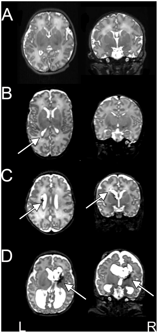
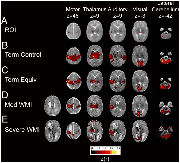
The cerebral white matter is vulnerable to injury in very preterm infants (born prior to 30 weeks gestation), resulting in a spectrum of lesions. These range from severe forms, including cystic periventricular leukomalacia and periventricular hemorrhagic infarction, to minor focal punctate lesions. Moderate to severe white matter injury in preterm infants has been shown to predict later neurodevelopmental disability, although outcomes can vary widely in infants with qualitatively comparable lesions. Resting state functional connectivity magnetic resonance imaging has been increasingly utilized in neurodevelopmental investigations and may provide complementary information regarding the impact of white matter injury on the developing brain. We performed resting state functional connectivity magnetic resonance imaging at term equivalent postmenstrual age in fourteen preterm infants with moderate to severe white matter injury secondary to periventricular hemorrhagic infarction. In these subjects, resting state networks were identifiable throughout the brain. Patterns of aberrant functional connectivity were observed and depended upon injury severity. Comparisons were performed against data obtained from prematurely-born infants with mild white matter injury and healthy, term-born infants and demonstrated group differences. These results reveal structural-functional correlates of preterm white matter injury and carry implications for future investigations of neurodevelopmental disability.


Conflict of interest statement

Competing Interests: The authors have declared that no competing interests exist.

Figures

Figure 1
Figure 1. WMI in preterm infants scanned at term equivalent PMA.
Transverse and coronal T2-weighted MR images illustrating (B) mild, (C) moderate and (D) severe WMI. Images from a healthy, term-born subject are provided for comparison (A). The arrows denote representative regions of injury. Areas of hemorrhage appear dark.
Figure 2
Figure 2. Neural network development in preterm infants with WMI scanned at term equivalent PMA.
Individual rs-fcMRI correlation maps illustrating Fisher z-transformed correlation coefficients (z(r); color threshold value = 0.3) overlaid on the subject-specific, atlas-registered T2-weighted images. Images include (A) seed ROIs in the motor cortex, thalamus, auditory and visual cortices and lateral cerebellum overlaid on population-specific atlas template; (D) moderate and (E) severe WMI; (B) healthy, term-born subject and (C) preterm infant with mild WMI provided for comparison. For moderate-severe WMI subjects, results were generated using an ROI located in the hemisphere of greater injury. Note incomplete RSN development most prominent in the hemisphere of greater injury (always shown on figure left).
Figure 3
Figure 3. Correlation matrices.
Matrices illustrating group mean Fisher z-transformed correlation coefficients for selected ROI pairs for (A) WMI, (B) term equivalent and (C) term control subjects. Also included are (D) term control – WMI, (E) term equivalent – WMI and (F) term control – term equivalent difference results. Note the lower magnitude correlation coefficients (positive as well as negative) in the WMI group in comparison to both the term equivalent and term control subjects. Black stars on matrices DF denote cells with between group differences on two-sample, two-tailed t-test (p<0.05; multiple comparisons correction not performed).
Figure 4
Figure 4. Severity of WMI correlates with loss of functional connectivity.
Scatter plots demonstrating the relationship between WMI score and Fisher z-transformed correlation coefficients evaluated in homologous ROI pairs for the (A) motor cortex and (B) thalamus for WMI subjects (blue diamonds). Lines illustrate the results of z(r) on WMI score linear regression. Correlation values with significance measures included. Results for term control subjects are displayed for comparison (red circles). All term control subjects had WMI scores of 0. Symbol abscissae have been shifted to avoid overlap.
Figure 5
Figure 5. RSN topography in relation to focal injury.
Fisher z-transformed correlation coefficient maps (color threshold value = 0.3) obtained with seed ROIs (illustrated in left column) overlaid on subject-specific, atlas-registered T2-weighted images. Results included from thalamic seeds in hemisphere of greater injury for infants with increasing WMI scores (scores 6, 10 and 12). Arrows denote areas of injury. Note the consistent preservation of functional connectivity in gray matter abutting the lesion and presence of functional connectivity in the hemisphere contralateral to the side of greatest injury.
Figure 6
Figure 6. Intrinsic BOLD signal fluctuations in WMI.
(A) T2-weighted image in an infant with severe WMI. (B) Voxelwise root mean squared temporal variance (signal SD; color threshold value = 0.15%). Note the paucity of activity in CSF spaces and persistence of BOLD signal fluctuations in gray matter despite severe thalamic injury. Resting state BOLD SD summed over (C) cerebral and (D) cerebellar ROIs in WMI and control infants. Each bar represents a single subject. Note the reduction of BOLD SD between the WMI (mean 1.73%) and control (mean 2.35%) groups for non-cerebellar ROIs (p<0.001) in (C). Values for cerebellar ROIs did not differ between WMI (mean 0.97%) and control (mean 1.06%) subjects (p = 0.36) as illustrated in (D). N.B., Values in panels B, C and D are computed relative to the whole brain mode value evaluated over all voxels and frames of each bold run.

References

    1. Adams-Chapman I, Hansen NI, Stoll BJ, Higgins R (2008) Neurodevelopmental outcome of extremely low birth weight infants with posthemorrhagic hydrocephalus requiring shunt insertion. Pediatrics 121: e1167–1177. - PMC - PubMed
    1. McCrea HJ, Ment LR (2008) The diagnosis, management, and postnatal prevention of intraventricular hemorrhage in the preterm neonate. Clin Perinatol 35: 777–792, vii. - PMC - PubMed
    1. Khwaja O, Volpe JJ (2008) Pathogenesis of cerebral white matter injury of prematurity. Arch Dis Child Fetal Neonatal Ed 93: F153–161. - PMC - PubMed
    1. Volpe JJ (2009) Brain injury in premature infants: a complex amalgam of destructive and developmental disturbances. Lancet Neurol 8: 110–124. - PMC - PubMed
    1. Pierson CR, Folkerth RD, Billiards SS, Trachtenberg FL, Drinkwater ME, et al. (2007) Gray matter injury associated with periventricular leukomalacia in the premature infant. Acta Neuropathol 114: 619–631. - PMC - PubMed

Publication types