. 2013 Jul 11;8(7):e68117.
doi: 10.1371/journal.pone.0068117. Print 2013.

Desmoglein-2-integrin Beta-8 interaction regulates actin assembly in endothelial cells: deregulation in systemic sclerosis

Affiliations

Desmoglein-2-integrin Beta-8 interaction regulates actin assembly in endothelial cells: deregulation in systemic sclerosis

Betti Giusti et al. PLoS One. .

Erratum in

  • PLoS One. 2013;8(7). doi:10.1371/annotation/b41766f2-c23d-455e-8d6e-e4bce5ae1d80

Abstract

Background: The inability of endothelial cells of patients affected by the diffuse form of Systemic sclerosis (SSc) to perform angiogenesis is a marker of the disease. We previously demonstrated that desmoglein-2 reduction is a major difference between (SSc)-microvascular endothelial cells (MVECs) and normal (N)-MVECs. Here we investigated the role of desmoglein-2 in human N-MVECs and SSc-MVECs angiogenesis.

Methodology/principal findings: Angiogenesis was studied by Matrigel invasion, capillary morphogenesis in vitro and Matrigel plug assay in vivo. Gene profiling was studied by Affymetrix technology and signal transduction by Western blotting. Colocalization was validated by immunoprecipitation and confocal microscopy. SiRNAs were used to validate the roles of specific molecules. We observed that desmoglein-2 co-localizes with integrin-beta8 in N-MVECs. This complex is required to signal through Rac, FAK, SMAD1/5 and MAP-kinases, promoting an angiogenic program. Inhibition of desmoglein-2 by DSG2-siRNA impaired actin stress fibres formation, capillary morphogenesis in vitro and angiogenesis in vivo. Transcriptome profiling after DSG2 inhibition revealed alterations of several genes involved in actin organization. siRNA inhibition of integrin-beta8 and RAC2 also resulted into capillary morphogenesis impairment in N-MVECs, due to reduced expression of the same actin-assembly genes that were down-regulated by DSG2 silencing. SSc-MVECs showed down-regulation of the same genes in DSG2-siRNA treated N-MVECs, suggesting that impairment of desmoglein-2/integrin-beta8 complex contributes to angiogenesis derangement in SSc. Transfection of DSG2 in SSc-MVEC partially restored their angiogenic properties in vitro.

Conclusions/significance: We have shown that impairment of actin assembly as a result of desmoglein-2/integrin-beta8 complex formation is a major factor contributing to angiogenesis deregulation in Systemic sclerosis.


Conflict of interest statement

Competing Interests: The authors have declared that no competing interests exist.

Figures

Figure 1
Figure 1. Effects of DSG2 silencing on in vitro parameters of angiogenesis of N-MVECs and in vivo Matrigel sponge assay.
A) DSG2 RT-PCR after DSG2 silencing. Data are expressed as fold change (siCONTROL-N-MVEC = 1). B) Representative desmoglein-2 Western blotting after DSG2 silencing. Results shown are representative of similar data obtained in 3 different experiments; molecular weights markers are to the right; beta-actin: loading control. The blot on the right refers to desmoglein-2 expression in three different SSc-MVEC lines (5, 6, and 7) used in the present study. C) Cell viability evaluated by WST-1 assay, D) proliferation and E) matrigel invasion in DSG2 silenced and control N-MVECs. Results are the mean ± SD of three different experiments performed in triplicate. *p<0.05, significantly different from control. F) Capillary morphogenesis at 6 h after seeding in Matrigel of control and treated N-MVECs. Numbers: percent field occupancy, taking control as 100%. Insets: morphology 24 hours after seeding. Data are from 3 experiments performed in triplicate. G) siDSG2-inhibition of angiogenesis in the Matrigel sponge model in mice. Upper part: angiogenesis quantification by haemoglobin content of each sponge. Pictures: ZEISS SR stereomicroscope aspect of the sponges under conditions corresponding to the histograms. Graphs are mean ± SD; * p<0.05. Results are the mean of three experiments (one animal for each condition, four Matrigel sponges in each animal).
Figure 2
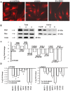
Figure 2. Effects of siDSG2 silencing in N-MVECs on stress fibres assembly and Rho/Rac transduction.
A) Actin stress fibers revealed by labeled phalloidin with a Nikon Plan Apo X60-oil immersion objective. ImageJ 1.44 software was used for image acquisition. 94±4% of siCONTROL-treated N-MVECs showed actin-stress fibers organization, while 89±11% siDSG2-treated N-MVECs and 100% SSc-MVECs exhibited a complete absence of actin stress fibers. B) Rho/Rac activation. Lanes 1, 2 and 3: total Rac and Rho in N-MVECs treated with control non-targeting siRNA, anti-DSG2 siRNA, and in SSc-MVECs, respectively; +GTP: positive control in N-MVECs lysate. Lanes 4, 5 and 6: constitutive Rho/Rac activation in control siRNA-N-MVECs, anti-DSG2 siRNA-N-MVECs and in SSc-MVECs, respectively. Actin: loading control. Position of molecular weight markers in kDa is shown on the right. A representative result of a single N-MVEC and SSc-MVEC line is shown. Similar results were obtained with all the N-MVEC and SSc-MVEC lines used in this study. C) RT-PCR validation of selected genes following DSG2 silencing in the three N-MVEC lines. D) Expression of the selected genes after ITGB8 and RAC2 silencing in N-MVECs. Results were normalized to GAPDH and fold changes of silenced-N-MVECs were calculated relative to the N-MVECs treated with control not-targeting siRNA. Data are expressed as log2 of fold change.
Figure 3
Figure 3. Colocalization of desmoglein-2 and integrin-beta8 in N-MVECs.
A) Immunostaining of N-MVECs with anti-desmoglein-2 and anti-integrin-beta8 antibodies (nuclei stained with DAPI) in a single N-MVEC (upper row) and in a group of N-MVECs (lower row). The results shown are indicative of experiments performed on 3 different N-MVEC cell lines. Original magnification: ×60 (Bio-Rad MRC 1024 ES Confocal Laser Scanning Microscope). B) Co-immunoprecipitation of integrin-beta8 and desmoglein-2. The lanes 1, 2 and 3 of each blot represent 1) input: total lysate, 2) protein A beads alone, 3) protein A beads + anti-Desmoglein-2 or anti-Integrin-beta8 antibody, respectively. Left side: immuno-blotting with anti-desmoglein-2 (upper part) and anti-integrin-beta8 (lower part) antibodies of the immuno-precipitates obtained with the same antibodies. Right side: immuno-blotting with anti-integrin-beta8 antibodies of the immuno-precipitate obtained with anti-desmoglein-2 antibodies (upper part), and immuno-blotting with anti-desmoglein-2 antibodies of the immuno-precipitate obtained with anti-integrin-beta8 antibodies (lower part). Numbers on the left indicate molecular weight (kDa) of the revealed bands. IP: immuno-precipitate; IB: immuno-blotting. The data shown represent a typical experiment out of three experiments on each N-MVEC line, all of which gave similar results.
Figure 4
Figure 4. Integrin-dependent and integrin-independent transduction pathways in N-MVECs and their modulation by silencing of DSG2 and ITGβ8.
Panel A: Western blotting. In each Western blot lane 1 indicates control conditions (1), lane 2 the effect of siDSG2 (2), and lane 3 the effect of siITGβ8 (3). Position of molecular weight markers in kDa is shown on the right. In each blot the non-phosphorylated form of the molecule is used as a loading control. The results shown have been selected among three different series of experiments that gave similar results in three different N-MVEC cell lines. For quantification, each electrophoresis pattern was subjected to densitometry. Histograms represent the average values ± SD of three different experiments performed in three different N-MVECs cell lines. *p<0.05, significantly different from control. Panel B: TRITC-labeled phalloidin immunofluorescence of stress fibers in control and in N-MVECs treated overnight with 5 µM FAK inhibitor 14, 30 µM MEK inhibitor UO126, 300 nM p38 inhibitor SB 202190, 10vµM TGFβRI/II inhibitor LY2109761.
Figure 5
Figure 5. Effects of ITGB8 silencing in N-MVECs.
Panel A. Capillary morphogenesis following ITGB8 silencing and relative controls. Pictures show the results of a typical experiment out of 3 experiments performed in triplicate with each N-MVEC line. Panel B: organization of actin stress fibers under the same conditions of A. Untreated N-MVECs showed actin stress fibers organization in 100% cells, siCONTROL-treated N-MVECs showed 93±6% of actin stress fibers-positive cells, while in siITGB8-treated N-MVECs actin stress fibers organization was absent in 91±8% cells.
Figure 6
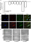
Figure 6. Real time PCR in SSc-MVECs of MACF, DIAPH1, DIAPH2, ARPC3, RAC2, CDH5, and ITGB8.
Rescue of the angiogenic phenotype in SSc-MVECs by transient transfection. Panel A: RT-PCR of the relevant transcripts in SSc-MVECs, relative to N-MVECs. Data are expressed as log2 of fold change. Negative values indicate decreased expression levels. Panel B: Immunostaining of SSc-MVECs with anti-desmoglein-2 and anti-integrin-beta8 antibodies (nuclei stained with DAPI) after transient transfection with control vector (upper pictures) or DSG2 transfection (lower pictures). The results shown are indicative of experiments performed on 3 SSc-MVEC cell lines. Original magnification: X60 (Bio-Rad MRC 1024 ES Confocal Laser Scanning Microscope). Panel C: Capillary morphogenesis in control and DSG2-transfected SSc-MVECs. Pictures show the results of a typical experiment out of 3 experiments performed in triplicate in each one of the different SSc-MVEC lines (SSc-MVEC 1, 2 and 3).

Similar articles

Cited by

References

    1. D'Alessio S, Fibbi G, Cinelli M, Guiducci S, Del Rosso A, et al. (2004) Matrix metalloproteinase 12-dependent cleavage of urokinase receptor in systemic sclerosis microvascular endothelial cells results in impaired angiogenesis. Arthritis Rheum 50: 3275–3285. - PubMed
    1. Margheri F, Manetti M, Serratì S, Nosi D, Pucci M, et al. (2006) Domain 1 of the urokinase-type plasminogen activator receptor is required for its morphologic and functional, beta2 integrin-mediated connection with actin cytoskeleton in human microvascular endothelial cells: failure of association in systemic sclerosis endothelial cells. Arthritis Rheum 54: 3926–3398. - PubMed
    1. Giusti B, Fibbi G, Margheri F, Serratì S, Rossi L, et al... (2006) A model of anti-angiogenesis: differential transcriptosome profiling of microvascular endothelial cells from diffuse systemic sclerosis patients. Arthritis Res Ther 8, R115. - PMC - PubMed
    1. Giusti B, Serratì S, Margheri F, Papucci L, Rossi L, et al. (2005) The antiangiogenic tissue kallikrein pattern of endothelial cells in systemic sclerosis. Arthritis Rheum 52: 3618–3628. - PubMed
    1. Suda K, Rothen-Rutishauser B, Günthert M, Wunderli-Allenspach H (2001) Phenotypic characterization of human umbilical vein endothelial (ECV304) and urinary carcinoma (T24) cells: endothelial versus epithelial features. In Vitro Cell Dev Biol Anim 37: 505–514. - PubMed

Publication types

MeSH terms