. 2013 Jul 9;8(7):e68624.
doi: 10.1371/journal.pone.0068624. Print 2013.

Diversity in protein profiles of individual calcium oxalate kidney stones

Affiliations

Diversity in protein profiles of individual calcium oxalate kidney stones

Nobuaki Okumura et al. PLoS One. .

Abstract

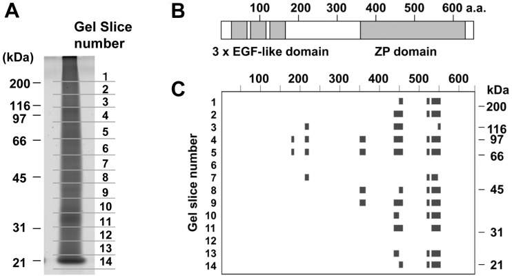
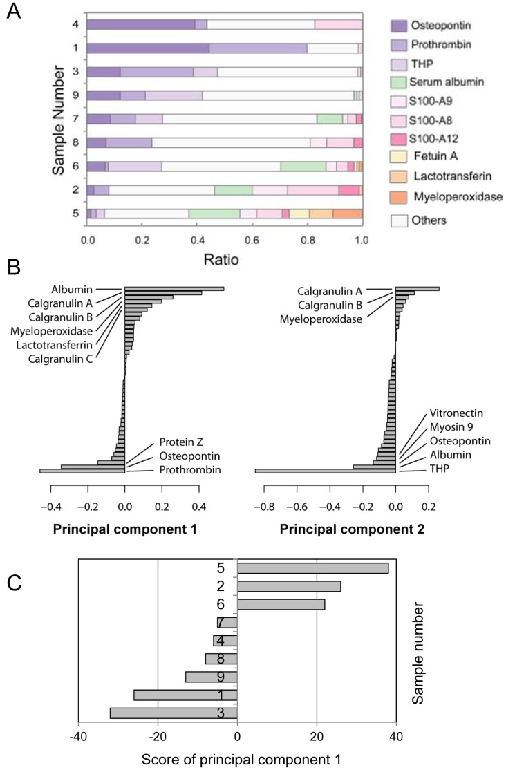
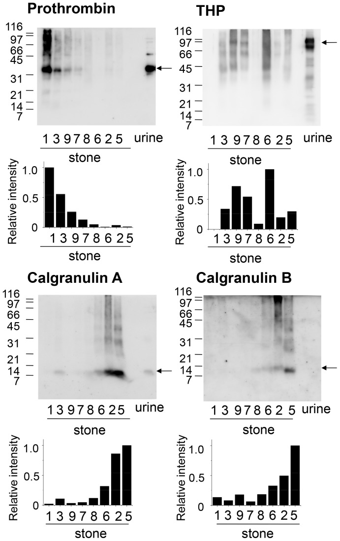
Calcium oxalate kidney stones contain low amounts of proteins, some of which have been implicated in progression or prevention of kidney stone formation. To gain insights into the pathophysiology of urolithiasis, we have characterized protein components of calcium oxalate kidney stones by proteomic approaches. Proteins extracted from kidney stones showed highly heterogeneous migration patterns in gel electrophoresis as reported. This was likely to be mainly due to proteolytic degradation and protein-protein crosslinking of Tamm-Horsfall protein and prothrombin. Protein profiles of calcium oxalate kidney stones were obtained by in-solution protease digestion followed by nanoLC-MALDI-tandem mass spectrometry, which resulted in identification of a total of 92 proteins in stones from 9 urolithiasis patients. Further analysis showed that protein species and their relative amounts were highly variable among individual stones. Although proteins such as prothrombin, osteopontin, calgranulin A and calgranulin B were found in most stones tested, some samples had high contents of prothrombin and osteopontin, while others had high contents of calgranulins. In addition, calgranulin-rich stones had various neutrophil-enriched proteins such as myeloperoxidase and lactotransferrin. These proteomic profiles of individual kidney stones suggest that multiple systems composed of different groups of proteins including leucocyte-derived ones are differently involved in pathogenesis of individual kidney stones depending on situations.


Conflict of interest statement

Competing Interests: The authors have declared that no competing interests exist.

Figures

Figure 1
Figure 1. Analysis of proteins extracted from CaOx kidney stones by SDS-PAGE.
CaOx kidney stones obtained from 8 individuals were solubilized, dialyzed, concentrated, and then the extracts containing 0.2 µg protein each were subjected to SDS-PAGE. Proteins were visualized by silver staining. Sample 4, the yield of which was <0.1 µg/mg stone, also showed similar diffused protein pattern (data not shown). Electrophoresis and staining were carried out three times, and all gave similar results.
Figure 2
Figure 2. Characterization of gel-separated proteins from a CaOx kidney stone by nanoLC MALDI-MS/MS analysis.
(A) One of the kidney stone extracts, Sample 3, was separated by SDS-PAGE, stained with Sypro Ruby, and cut into 14 pieces as indicated. Each gel piece was subjected to in gel trypsin digestion, and the resultant peptides were analyzed by nanoLC/MALDI MS/MS followed by database search on MASCOT. The results were presented as Table 2. (B) Domain structure of human THP (SwissProt : UROM_HUMAN). (C) From the results shown in Table 2, peptides matched to THP were mapped on the amino acid sequence of human THP.
Figure 3
Figure 3. Procedure for protein profiling of CaOx renal stones.
In order to analyze the protein compositions of individual CaOx stones, extracts from CaOx renal stones were subjected to protease digestion in solution and analyzed using nano-LC and MALDI-MS/MS as summarized in this scheme.
Figure 4
Figure 4. Classification of protein components detected in CaOx kidney stones.
Extracts from 9 individual CaOx stone samples were examined separately by shotgun analysis as shown in Fig. 3, and the results in detail were presented in Table S1. As summarized in this figure, a total of 92 proteins were identified in this analysis. These proteins could be classified into three major groups according to their possible origins, i. e., kidney proteins, blood proteins and leucocyte-related proteins. Proteins in each group were further classified into subgroups.
Figure 5
Figure 5. Comparison of relative peptide numbers detected of individual CaOx kidney stones.
(A) After nano-LC/MALDI-MS/MS analysis, the numbers of peptide hits were presented as a bar graph with the total number of peptide hits in each gel sample as 100%. (B) The numbers of peptide hits were subjected to principal component analysis, and the first (left) and the second (right) eigenvectors were shown in a decreasing order. (C) The score of principal component 1 of each sample was presented as a bar graph.
Figure 6
Figure 6. Western blot analysis of major proteins in CaOx kidney stones.
Extracts of CaOx kidney stones containing 0.1 µg proteins were separated by SDS-PAGE and blotted on a PVDF membrane. To confirm the reactivity of antibodies and indicate the size of the proteins in urine, a concentrated urine sample from a normal subject (1 µg protein) was applied on the last lane of each gel. The membrane was then probed with anti-prothrombin fragment 1, anti-calgranulin A, anti-calgranulin B or anti-THP antibodies. Arrows indicate the positions of prothrombin fragment 1 (upper left), full length THP (upper right), full length calgranulin A (lower left), and full length calgranulin B (lower right). Western blotting was repeated two times and gave similar results. When Sample 1, 3, 9 and 7 (Group 1) was compared with Sample 8, 6, 2 and 5 (Group 2), p-value of Mann-Whitney U-test was 0.028 for prothrombin, calgranulin A and calgranulin B, while that for THP was 0.8857.

References

    1. Tsujihata M (2008) Mechanisms of calcium oxalate renal stone formation and renal tubular cell injury. Int J Urol 15: 115–120. - PubMed
    1. Robertson WG, Peacock M, Nordin BEC (1968) Activity products in stone-forming and non-stone forming urine. Clin Sci 34: 579–594. - PubMed
    1. Boyce WH (1968) Organic matrix of human urinary concretions. Am J Med 45: 673–683. - PubMed
    1. Maxfield M (1963) Urinary mucopolysaccharides and calculi. Ann Rev Med 14: 99–. - PubMed
    1. Kohri K, Nomura S, Kitamura Y, Nagata T, Yoshioka K, et al. (1993) Structure and expression of the mRNA encoding urinary stone protein (osteopontin). J Biol Chem 268: 15180–15184. - PubMed

Publication types

Supplementary concepts