. 2013 Jul 18;8(7):e68935.
doi: 10.1371/journal.pone.0068935. Print 2013.

The role of peroxisome proliferator-activated receptor and effects of its agonist, pioglitazone, on a rat model of optic nerve crush: PPARγ in retinal neuroprotection

Affiliations

The role of peroxisome proliferator-activated receptor and effects of its agonist, pioglitazone, on a rat model of optic nerve crush: PPARγ in retinal neuroprotection

Juming Zhu et al. PLoS One. .

Abstract

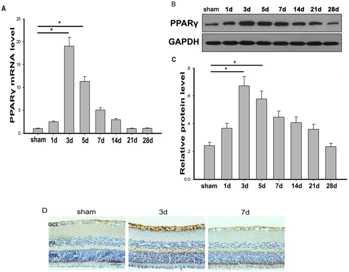
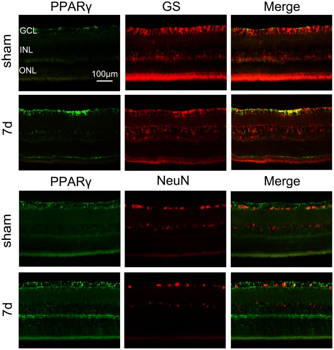
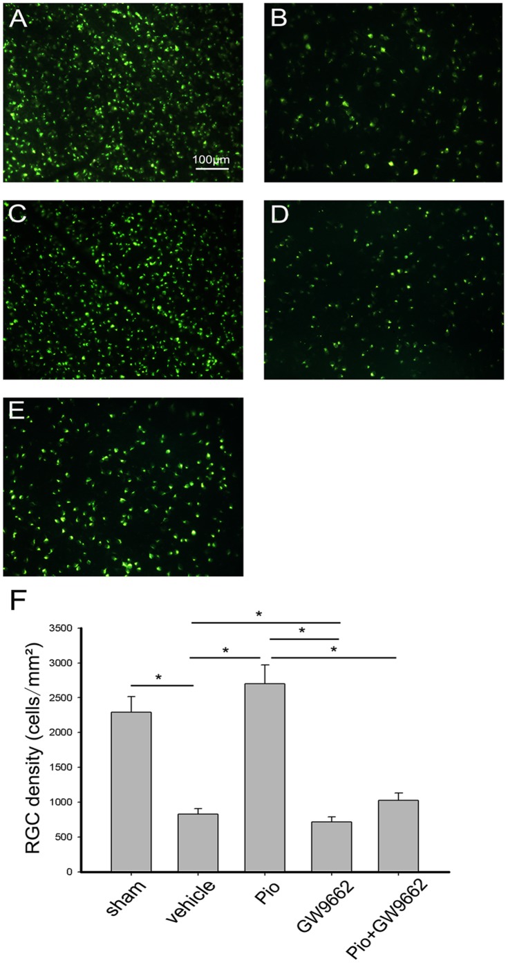
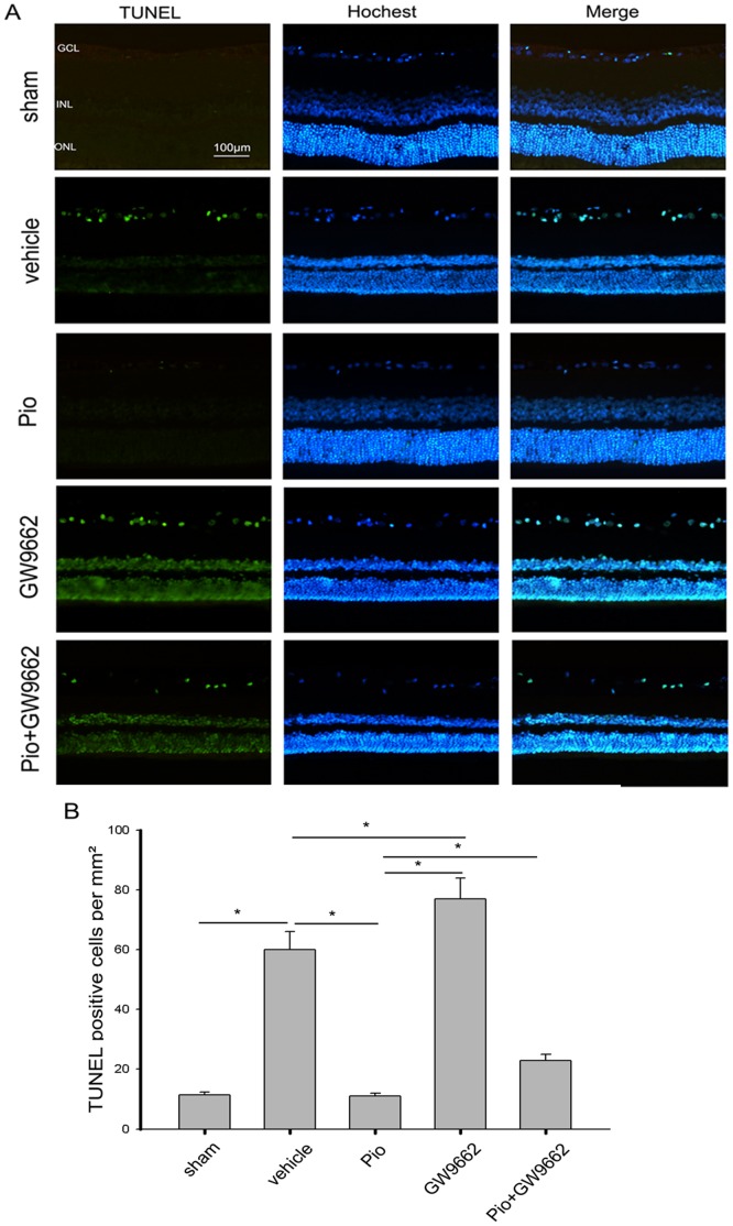
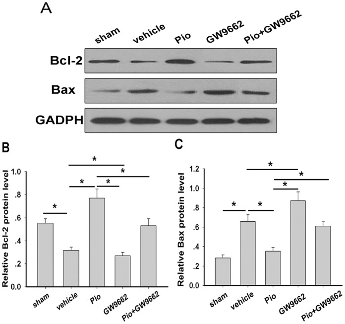
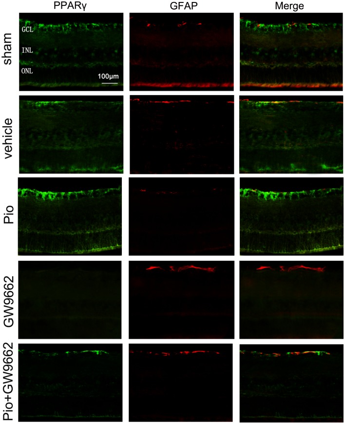
It has been shown that peroxisome proliferators-activated receptor gamma (PPARγ) is beneficial for central nervous system injury. However its role on optic nerve injury remains unknown. In the present study, we examined the change of PPARγ expression in rat retina following optic nerve injury and investigated the effect of pioglitazone (Pio), a PPARγ agonist, on retinal ganglion cells (RGCs) neuroprotection using a rat optic nerve crush (ONC) model. Our results showed that PPARγ mRNA and protein levels were increased after ONC, and most of PPARγ-immunoreactive cells colocalized with Müller cells. Pio treatment significantly enhanced the number of surviving RGCs and inhibited RGCs apoptosis induced by ONC. However, when PPARγ antagonist GW9662 was used, these neuroprotective effects were abolished. In addition, pio attenuated Müller cell activation after ONC. These results indicate that PPARγ appears to protect RGCs from ONC possibly via the reduction of Müller glial activation. It provides evidence that activation of PPARγ may be a potential alternative treatment for RGCs neuroprotection.


Conflict of interest statement

Competing Interests: The authors have declared that no competing interests exist.

Figures

Figure 1
Figure 1. The expression of PPARγ in rat retina after ONC.
A. The expression of PPARγ mRNA was increased after ONC. The highest expression was found on day 3, then returned to basal values on day 14 and remained unchanged until the end of the observation period of 28 days(n = 6 in each group). *p<0.05. B. Western blot analysis of PPARγ protein in the retina. C. The expression of PPARγ protein relative to GAPDH was determined by measuring the optical density using ImageJ software(n = 6 in each group). *p<0.05. D. Immunohistochemical staining of PPARγ in rat retina. High PPARγ immunoreactivity was observed in the nerve fiber layer (NFL), ganglion cell layer (GCL), inner neuclear layer (INL) and outer plexiform layer (OPL) 3 days after ONC. Bar = 100 µm.
Figure 2
Figure 2. Double immunofluorescence staining for PPARγ (green)/GS (red) and PPARγ (green)/NeuN (red) on the normal retina and the retina 7 days after ONC.
Most of PPARγ positive cells are colocalized with GS positive Müller cells, not within NeuN positive RGCs. Bar = 100 µm.
Figure 3
Figure 3. Effect of pioglitazone (Pio) on RGCs survival after optic nerve crush (ONC).
(A) A representative photograph of RGCs labeled with FluoroGold (FG) injected into the superior colliculus in normal retina (sham group). (B–E) A representative photograph of FG-labeled RGCs, 7 days after ONC with intraperitoneal injection of DMSO(B), Pio(C), GW9662(D) and Pio+ GW9662(E). Bar = 100 µm. (F) Quantitative analysis of the number of FG-labeled RGCs 7 days after ONC. Data represent the means ± SD (n = 6 in each group). *p<0.05.
Figure 4
Figure 4. The anti-apoptotic effect of Pio on RGCs after ONC.
(A) Representative of the terminal dUTP nick end labeling (TUNEL) staining in retinal sections of each group at 7 days after ONC. Hochest dye was used for nuclear staining. (B) TUNEL-positive cells in the RGCs layer at 7 days after ONC(n = 6 in each group). *p<0.05. Bar = 100 µm.
Figure 5
Figure 5. Effect of Pio on anti- and pro- apoptotic protein expression in rat retina at 7 days after ONC.
Western blot analysis indicated that Pio treatment could increase the expression of Bcl-2 (A and B), and decrease the expression of Bax (A and C). Bars represent the means ± SD (n = 6 in each group). *p<0.05.
Figure 6
Figure 6. Effect of Pio on Müller cell activation in retina.
Immunoreactivity for PPARγ (green) and GFAP (red) in rat retinal sections of each group at 7days after ONC. Bar = 100 µm.

Similar articles

Cited by

References

    1. Schmidt KG, Bergert H, Funk RH (2008) Neurodegenerative diseases of the retina and potential for protection and recovery. Curr Neuropharmacol 6: 164–178. - PMC - PubMed
    1. Kisiswa L, Dervan AG, Albon J, Morgan JE, Wride MA (2010) Retinal ganglion cell death postponed: giving apoptosis a break? Ophthalmic Res 43: 61–78. - PubMed
    1. Quigley HA, McKinnon SJ, Zack DJ, Pease ME, Kerrigan-Baumrind LA, et al. (2000) Retrograde axonal transport of BDNF in retinal ganglion cells is blocked by acute IOP elevation in rats. Invest Ophthalmol Vis Sci 41: 3460–3466. - PubMed
    1. Sucher NJ, Lipton SA, Dreyer EB (1997) Molecular basis of glutamate toxicity in retinal ganglion cells. Vision Res 37: 3483–3493. - PubMed
    1. Tezel G (2006) Oxidative stress in glaucomatous neurodegeneration: mechanisms and consequences. Prog Retin Eye Res 25: 490–513. - PMC - PubMed

Publication types

MeSH terms