

Activation of Egr-1 in human lung epithelial cells exposed to silica through MAPKs signaling pathways
- PMID: 23874821
- PMCID: PMC3715534
- DOI: 10.1371/journal.pone.0068943
Activation of Egr-1 in human lung epithelial cells exposed to silica through MAPKs signaling pathways
Abstract
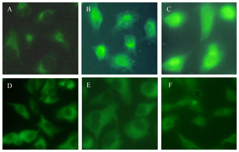
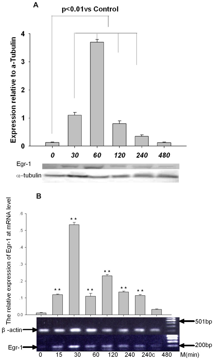
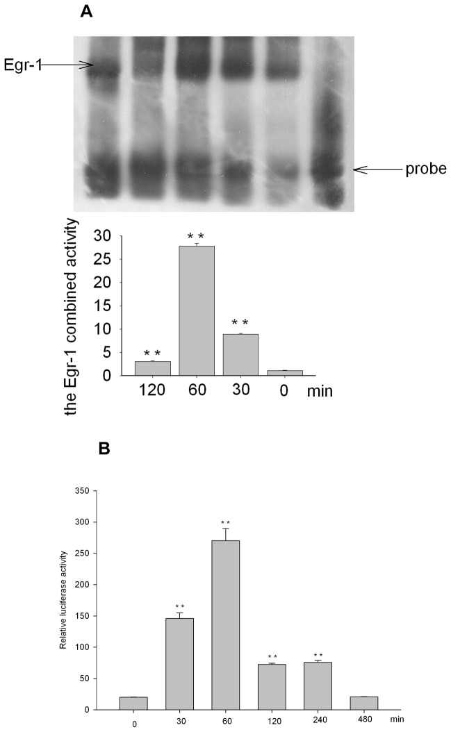
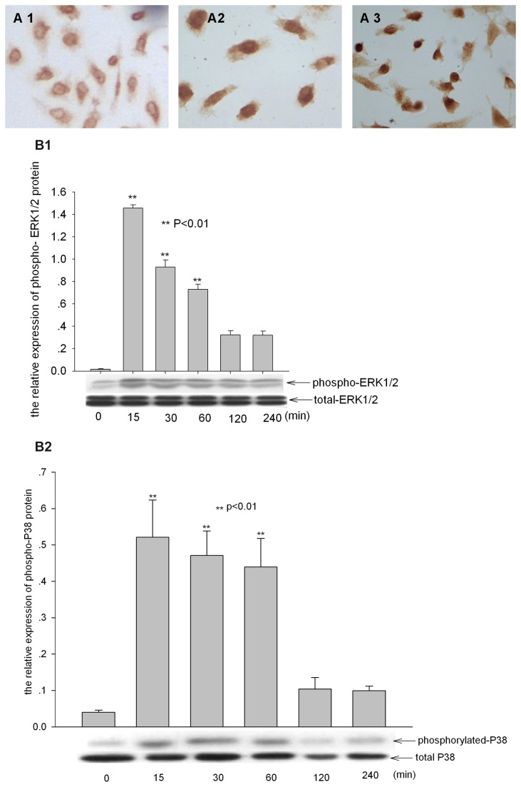
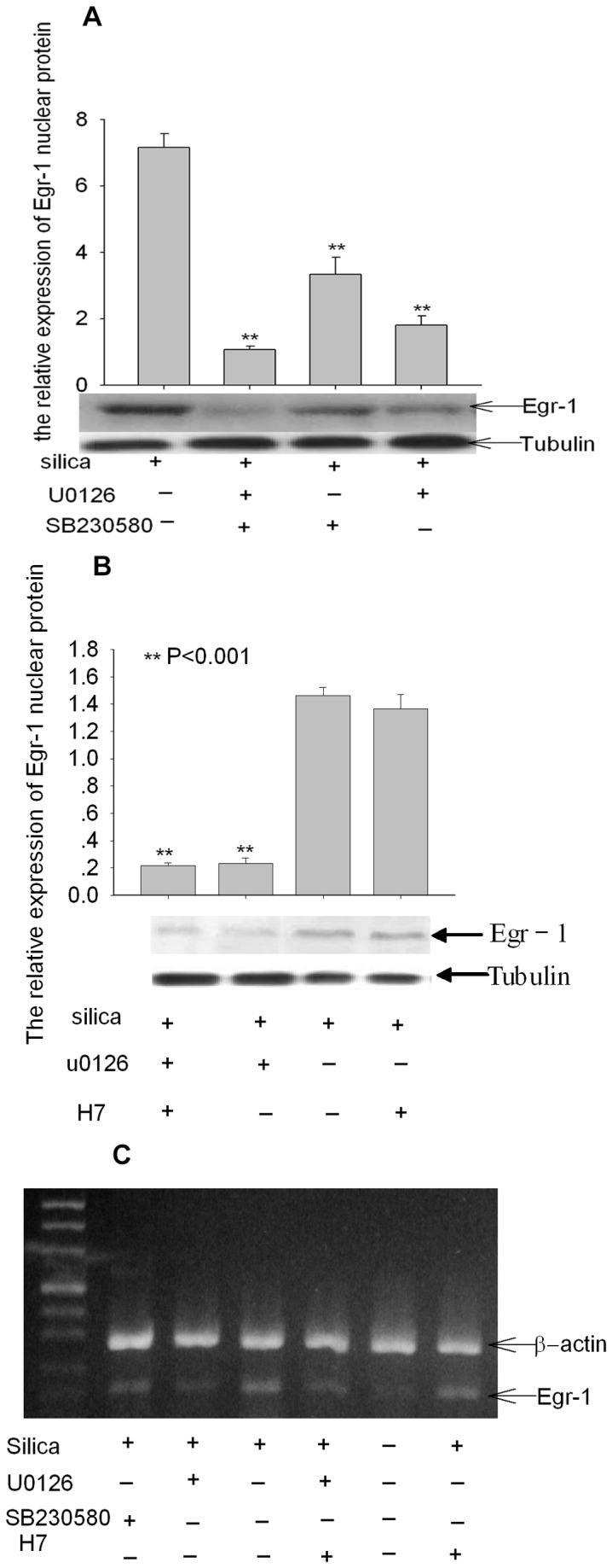
The alveolar type II epithelial cell, regarded historically as a key target cell in initial injury by silica, now appears to be important in both defense from lung damage as well as elaboration of chemokines and cytokines. The molecular basis for silica-induced epithelial cell injury is poorly understood. In this study we explored the activation of nuclear factor Egr-1 and related signal pathway. Human II alveolar epithelial line A549 cells were exposed to silica for indicated time to assay the expression and activation of Egr-1 and upstream MAPKs. Immunofluorescence, western-blot techniques, RT-PCR, Electrophoretic mobility shift assay (EMSA), transient transfection assay, kinase inhibitor experiments were performed. It was found that the expression of Egr-1 at mRNA and protein level was significantly increased in A549 cells after administration with silica and the activity of Egr-1 peaked by silica treatment for 60 minutes. Furthermore, phosphorylated-ERK1/2, P38 MAPKs (the upstream kinase of Egr-1) ballooned during 15-30minutes, 30-60minutes respectively after silica exposure in A549 cells. By administration of ERK1/2, P38 inhibitor, the expression and transcription of Egr-1 were both markedly decreased. But PKC inhibitor did not prevent the increase of Egr-1. These results indicated Egr-1 played a critical role in silica-induced pulmonary fibrosis in an ERK1/2, P38 MAPKs-dependent manner, which suggests Egr-1 is an essential regulator in silicosis, and underlines a new molecular mechanism for fibrosis induced by silica.
Conflict of interest statement
Figures
Similar articles
-
p38 and Src-ERK1/2 pathways regulate crystalline silica-induced chemokine release in pulmonary epithelial cells.Toxicol Sci. 2004 Oct;81(2):480-90. doi: 10.1093/toxsci/kfh214. Epub 2004 Jul 7. Toxicol Sci. 2004. PMID: 15240896
-
[Sudy on the activation of early growth response factor-1 by silica dioxide and its signal pathway].Zhonghua Bing Li Xue Za Zhi. 2005 May;34(5):293-6. Zhonghua Bing Li Xue Za Zhi. 2005. PMID: 16181552 Chinese.
-
[Expression of early growth response gene-1 in macrophages stimulated by silicon dioxide].Zhonghua Bing Li Xue Za Zhi. 2003 Dec;32(6):558-62. Zhonghua Bing Li Xue Za Zhi. 2003. PMID: 14761604 Chinese.
-
Up-regulation of early growth response gene 1 (EGR-1) via ERK1/2 signals attenuates sulindac sulfide-mediated cytotoxicity in the human intestinal epithelial cells.Toxicol Appl Pharmacol. 2007 Sep 1;223(2):155-63. doi: 10.1016/j.taap.2007.04.018. Epub 2007 May 21. Toxicol Appl Pharmacol. 2007. PMID: 17599376
-
PKCδ-dependent activation of ERK1/2 leads to upregulation of the human NHE2 transcriptional activity in intestinal epithelial cell line C2BBe1.Am J Physiol Gastrointest Liver Physiol. 2012 Feb 1;302(3):G317-25. doi: 10.1152/ajpgi.00363.2011. Epub 2011 Nov 3. Am J Physiol Gastrointest Liver Physiol. 2012. PMID: 22052014 Free PMC article.
Cited by
-
Targeted migration of bone marrow mesenchymal stem cells inhibits silica-induced pulmonary fibrosis in rats.Stem Cell Res Ther. 2018 Dec 4;9(1):335. doi: 10.1186/s13287-018-1083-y. Stem Cell Res Ther. 2018. PMID: 30514375 Free PMC article.
-
P. aeruginosa type III and type VI secretion systems modulate early response gene expression in type II pneumocytes in vitro.BMC Genomics. 2022 May 4;23(1):345. doi: 10.1186/s12864-022-08554-0. BMC Genomics. 2022. PMID: 35508983 Free PMC article.
-
Effects of Estrogen and Estrogen Receptors on Transcriptomes of HepG2 Cells: A Preliminary Study Using RNA Sequencing.Int J Endocrinol. 2018 Oct 28;2018:5789127. doi: 10.1155/2018/5789127. eCollection 2018. Int J Endocrinol. 2018. PMID: 30510575 Free PMC article.
-
Genome-wide mRNA profiling identifies the NRF2-regulated lymphocyte oxidative stress status in patients with silicosis.J Occup Med Toxicol. 2021 Sep 13;16(1):40. doi: 10.1186/s12995-021-00332-0. J Occup Med Toxicol. 2021. PMID: 34517882 Free PMC article.
-
DT(270-326) , a Truncated Diphtheria Toxin, Increases Blood-Tumor Barrier Permeability by Upregulating the Expression of Caveolin-1.CNS Neurosci Ther. 2016 Jun;22(6):477-87. doi: 10.1111/cns.12519. Epub 2016 Feb 10. CNS Neurosci Ther. 2016. PMID: 26861687 Free PMC article.
References
-
- Lee CG, Cho SJ, Kang MJ, Chapoval SP, Lee PJ et al. (2004) Early growth response gene 1-mediated apoptosis is essential for transforming growth factor beta1-induced pulmonary fibrosis. J Exp Med 200: 377-389. doi:10.1084/jem.20040104. PubMed: 15289506. - DOI - PMC - PubMed
-
- Pritchard MT, Nagy LE (2005) Ethanol-induced liver injury: potential roles for egr-1. Alcohol Clin Exp Res 29: 146S-150S. doi:10.1097/01.alc.0000189286.81943.51. PubMed: 16344600. - DOI - PubMed
-
- Zhang Y, Bonzo JA, Gonzalez FJ, Wang L (2011) Diurnal regulation of the early growth response 1 (Egr-1) protein expression by hepatocyte nuclear factor 4alpha (HNF4alpha) and small heterodimer partner (SHP) cross-talk in liver fibrosis. J Biol Chem 286: 29635-29643. doi:10.1074/jbc.M111.253039. PubMed: 21725089. - DOI - PMC - PubMed
Publication types
MeSH terms
Substances
LinkOut - more resources
Full Text Sources
Other Literature Sources
Medical
Miscellaneous
