. 2013 Jul 9;8(7):e69003.
doi: 10.1371/journal.pone.0069003. Print 2013.

Structural analysis of mitochondrial mutations reveals a role for bigenomic protein interactions in human disease

Affiliations

Structural analysis of mitochondrial mutations reveals a role for bigenomic protein interactions in human disease

Rhiannon E Lloyd et al. PLoS One. .

Abstract

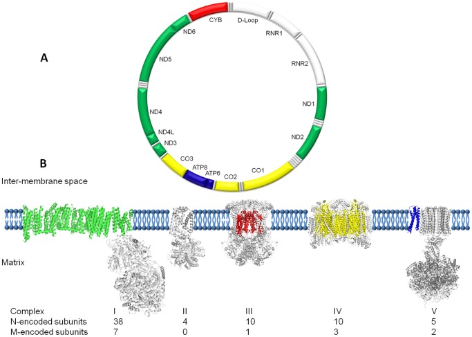
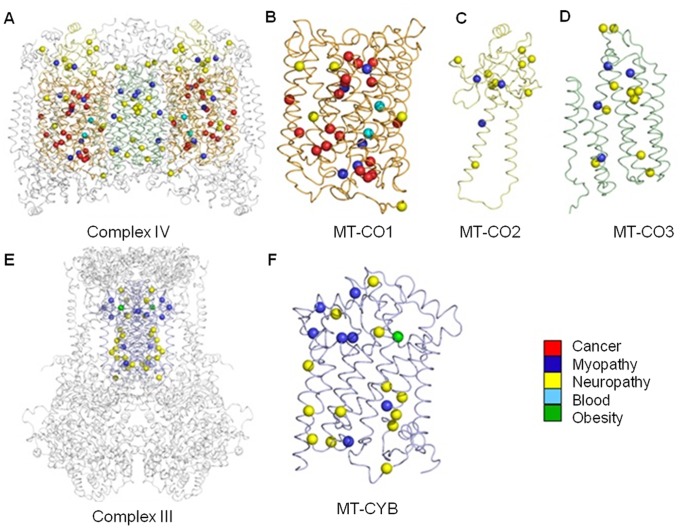
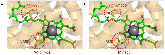
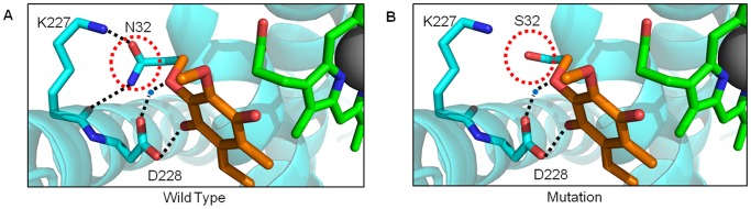
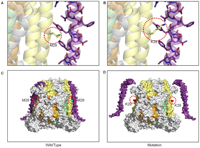
Mitochondria are the energy producing organelles of the cell, and mutations within their genome can cause numerous and often severe human diseases. At the heart of every mitochondrion is a set of five large multi-protein machines collectively known as the mitochondrial respiratory chain (MRC). This cellular machinery is central to several processes important for maintaining homeostasis within cells, including the production of ATP. The MRC is unique due to the bigenomic origin of its interacting proteins, which are encoded in the nucleus and mitochondria. It is this, in combination with the sheer number of protein-protein interactions that occur both within and between the MRC complexes, which makes the prediction of function and pathological outcome from primary sequence mutation data extremely challenging. Here we demonstrate how 3D structural analysis can be employed to predict the functional importance of mutations in mtDNA protein-coding genes. We mined the MITOMAP database and, utilizing the latest structural data, classified mutation sites based on their location within the MRC complexes III and IV. Using this approach, four structural classes of mutation were identified, including one underexplored class that interferes with nuclear-mitochondrial protein interactions. We demonstrate that this class currently eludes existing predictive approaches that do not take into account the quaternary structural organization inherent within and between the MRC complexes. The systematic and detailed structural analysis of disease-associated mutations in the mitochondrial Complex III and IV genes significantly enhances the predictive power of existing approaches and our understanding of how such mutations contribute to various pathologies. Given the general lack of any successful therapeutic approaches for disorders of the MRC, these findings may inform the development of new diagnostic and prognostic biomarkers, as well as new drugs and targets for gene therapy.


Conflict of interest statement

Competing Interests: The authors have declared that no competing interests exist.

Figures

Figure 1
Figure 1. Architecture of the mitochondrial genome and respiratory chain.
(A) Schematic representation of the 16,569 bp human mitochondrial genome (NC_012920), with the protein-coding genes colored according to the complexes to which they contribute subunits, two ribosomal RNAs, 22 tRNAs and non-coding D-loop in white. (B) Montage depicting the structural information currently available for the five complexes that together contribute to the mitochondrial oxidative phosphorylation machinery. Each complex (to scale) is embedded in a cartoon representation of the lipid bilayer with the mitochondrial (m)-encoded subunits colored corresponding to the genome diagram. The nuclear (n)-encoded subunits are shown in grey with the relative contributions found in higher organisms detailed below.
Figure 2
Figure 2. A new structural map of human mitochondrial disease mutation sites in Complexes III and IV.
Mitochondrial diseases result in diverse pathology, and can be multi-systemic or tissue-specific. Here we show 93 individual mutation sites mapped onto their corresponding 3D crystal structures. Each mutation site is shown as a sphere colored according to the primary pathology or tissue (see legend) found to be affected in either single or groups of patients. The complete dimeric Complex IV is depicted as a ribbon model (A) with the mitochondrial-encoded subunits, MT-CO1 (B), MT-CO2 (C) and MT-CO3 (D), colored orange, yellow and green, respectively. The three COX monomers are shown separately for clarity. The dimeric Complex III is shown in the same format (E) with the mitochondrial-encoded MT-CYB (F) subunits colored as blue ribbons.
Figure 3
Figure 3. Active site mutations.
(A) The active site region of the wild-type MT-CO1 subunit of complex IV is depicted as a ribbon diagram with key amino acids as orange stick representations (within red circles). The heme a 3 is colored in green with the Fe atom in grey. (B) Two separate mutations have been modeled, I280T and V380I. More detailed diagrams can be found in Figure S5.
Figure 4
Figure 4. The role of residues contributing to substrate-binding cavities.
MT-CYB has several deep pockets for binding, redox interaction and modification of substrates. (A) The Qi site is formed by the contribution of multiple helical regions (blue) that fold around heme b H (green) while maintaining contact with the solvent. The wild-type residue N32 (PDB 1NTZ) is shown making hydrogen bonds (dotted lines) with the natural substrate ubiquinone (orange), within the same pocket. (B) Mutation to S32 results in the loss of key hydrogen bonds previously identified to be crucial in the redox mechanism.
Figure 5
Figure 5. Assembly disruption from bigenomic protein incompatibility in Complex IV.
(A) The mitochondrial subunits are surrounded by nuclear-encoded subunits that make intimate interactions, in this case MT-CO2 (yellow ribbons) with COX6C (purple ribbon), respectively. The wild type M29 residue is shown in stick form occupying a position between to aromatic side chains from COX6C. (B) The mutation K29 results in the incorporation of a long and polar side chain that is incompatible within the tight interface between MT-CO2 and COX6C. (C) The wild type-enzyme is shown as a surface model in the same color scheme with the position of the mutation site highlighted by a dotted red circle. (D) A surface illustration depicts the position of the K29 mutation and the potential resulting aberrant assembly of the nuclear subunit COX6C.

Similar articles

Cited by

References

    1. Efremov RG, Sazanov LA (2011) Structure of the membrane domain of respiratory complex I. Nature. 476: 414–420. - PubMed
    1. Baradaran R, Berrisford JM, Minhas GS, Sazanov LA (2013) Crystal structure of the entire respiratory complex I. Nature. 494: 443–448. - PMC - PubMed
    1. Junge W, Sielaff H, Engelbrecht S (2009) Torque generation and elastic power transmission in the rotary F(O)F(1)-ATPase. Nature 459: 364–370. - PubMed
    1. Vafai SB, Mootha VK (2012) Mitochondrial disorders as windows into an ancient organelle. Nature 491: 374–383. - PubMed
    1. Hosler J (2012) Biogenesis/assembly of respiratory enzyme complexes. Biochim Biophys Acta 1817: 849–850. - PubMed

Publication types

MeSH terms

Substances

LinkOut - more resources