

Effect of epicatechin against radiation-induced oral mucositis: in vitro and in vivo study
- PMID: 23874895
- PMCID: PMC3715464
- DOI: 10.1371/journal.pone.0069151
Effect of epicatechin against radiation-induced oral mucositis: in vitro and in vivo study
Abstract
Purpose: Radiation-induced oral mucositis limits the delivery of high-dose radiation to head and neck cancer. This study investigated the effectiveness of epicatechin (EC), a component of green tea extracts, on radiation-induced oral mucositis in vitro and in vivo.
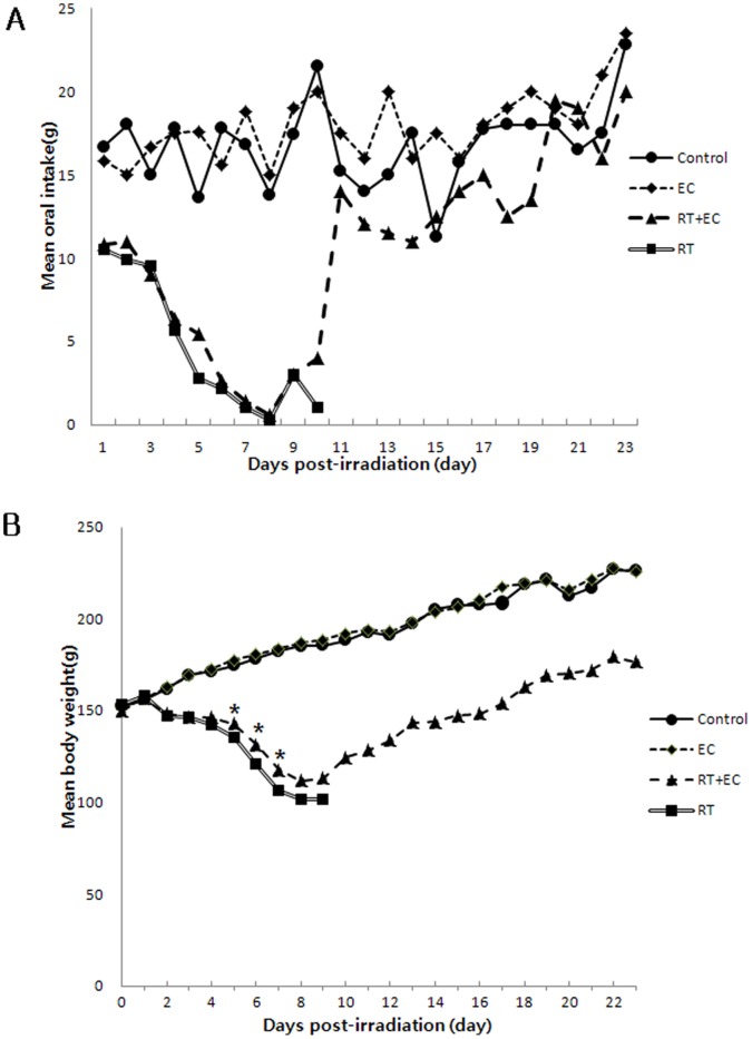
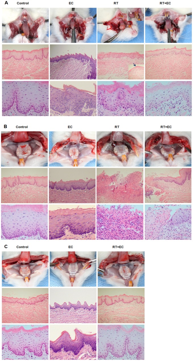
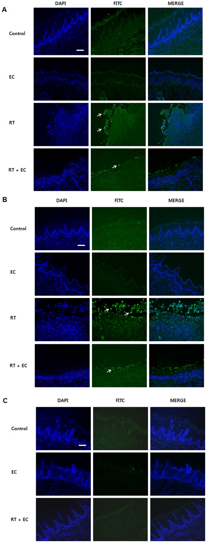
Experimental design: The effect of EC on radiation-induced cytotoxicity was analyzed in the human keratinocyte line HaCaT. Radiation-induced apoptosis, change in mitochondrial membrane potential (MMP), reactive oxygen species (ROS) generation and changes in the signaling pathway were investigated. In vivo therapeutic effects of EC for oral mucositis were explored in a rat model. Rats were monitored by daily inspections of the oral cavity, amount of oral intake, weight change and survival rate. For histopathologic evaluation, hematoxylin-eosin staining and TUNEL staining were performed.
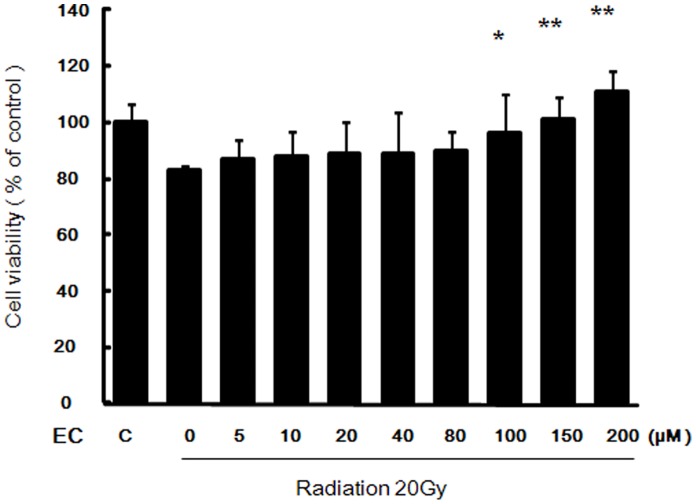
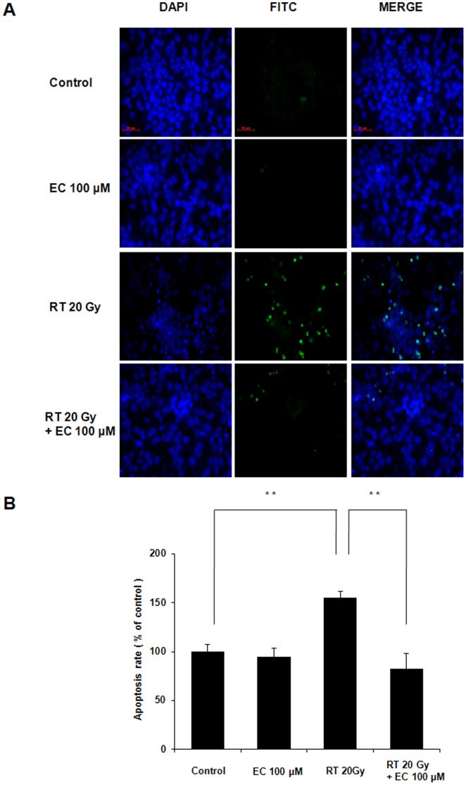
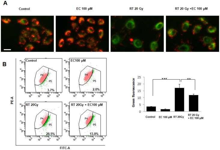
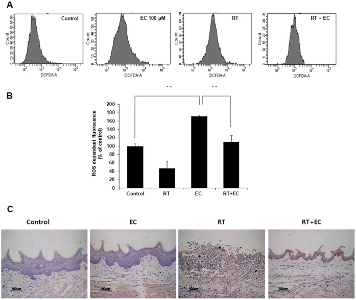
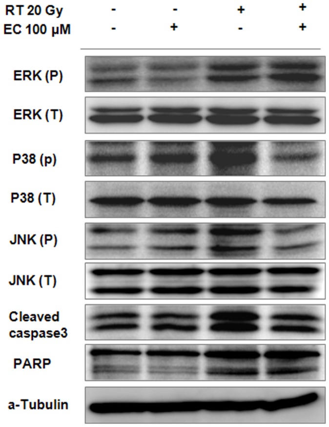
Results: EC significantly inhibited radiation-induced apoptosis, change of MMP, and intracellular ROS generation in HaCaT cells. EC treatment markedly attenuated the expression of p-JNK, p-38, and cleaved caspase-3 after irradiation in the HaCaT cells. Rats with radiation-induced oral mucositis showed decreased oral intake, weight and survival rate, but oral administration of EC significantly restored all three parameters. Histopathologic changes were significantly decreased in the EC-treated irradiated rats. TUNEL staining of rat oral mucosa revealed that EC treatment significantly decreased radiation-induced apoptotic cells.
Conclusions: This study suggests that EC significantly inhibited radiation-induced apoptosis in keratinocytes and rat oral mucosa and may be a safe and effective candidate treatment for the prevention of radiation-induced mucositis.
Conflict of interest statement
Figures
Similar articles
-
Epicatechin inhibits radiation-induced auditory cell death by suppression of reactive oxygen species generation.Neuroscience. 2011 Dec 29;199:410-20. doi: 10.1016/j.neuroscience.2011.09.012. Epub 2011 Sep 16. Neuroscience. 2011. PMID: 21946009
-
Protective Effects of N-Acetylcysteine against Radiation-Induced Oral Mucositis In Vitro and In Vivo.Cancer Res Treat. 2020 Oct;52(4):1019-1030. doi: 10.4143/crt.2020.012. Epub 2020 Jun 18. Cancer Res Treat. 2020. PMID: 32599978 Free PMC article.
-
Protective effects of Korean red ginseng against radiation-induced apoptosis in human HaCaT keratinocytes.J Radiat Res. 2014 Mar 1;55(2):245-56. doi: 10.1093/jrr/rrt109. Epub 2013 Sep 26. J Radiat Res. 2014. PMID: 24078877 Free PMC article.
-
Potential prevention: Aloe vera mouthwash may reduce radiation-induced oral mucositis in head and neck cancer patients.Chin J Integr Med. 2012 Aug;18(8):635-40. doi: 10.1007/s11655-012-1183-y. Epub 2012 Aug 2. Chin J Integr Med. 2012. PMID: 22855041 Review.
-
Antioxidant agents: a future alternative approach in the prevention and treatment of radiation-induced oral mucositis?Altern Ther Health Med. 2015 Mar-Apr;21(2):36-41. Altern Ther Health Med. 2015. PMID: 25830279 Review.
Cited by
-
Radiation-Induced Normal Tissue Damage: Oxidative Stress and Epigenetic Mechanisms.Oxid Med Cell Longev. 2019 Nov 12;2019:3010342. doi: 10.1155/2019/3010342. eCollection 2019. Oxid Med Cell Longev. 2019. PMID: 31781332 Free PMC article. Review.
-
Epicatechin stimulates mitochondrial activity and selectively sensitizes cancer cells to radiation.PLoS One. 2014 Feb 6;9(2):e88322. doi: 10.1371/journal.pone.0088322. eCollection 2014. PLoS One. 2014. PMID: 24516636 Free PMC article.
-
Molecular Mechanisms and Therapeutic Effects of (-)-Epicatechin and Other Polyphenols in Cancer, Inflammation, Diabetes, and Neurodegeneration.Oxid Med Cell Longev. 2015;2015:181260. doi: 10.1155/2015/181260. Epub 2015 Jun 9. Oxid Med Cell Longev. 2015. PMID: 26180580 Free PMC article. Review.
-
Reduction-oxidation (redox) system in radiation-induced normal tissue injury: molecular mechanisms and implications in radiation therapeutics.Clin Transl Oncol. 2018 Aug;20(8):975-988. doi: 10.1007/s12094-017-1828-6. Epub 2018 Jan 9. Clin Transl Oncol. 2018. PMID: 29318449 Review.
-
Protective Role of Natural Compounds under Radiation-Induced Injury.Nutrients. 2022 Dec 17;14(24):5374. doi: 10.3390/nu14245374. Nutrients. 2022. PMID: 36558533 Free PMC article. Review.
References
-
- Sankaranarayanan R, Masuyer E, Swaminathan R, Ferlay J, Whelan S (1998) Head and neck cancer: a global perspective on epidemiology and prognosis. Anticancer Res 18: 4779–4786. - PubMed
-
- Pignon JP, Bourhis J, Domenge C, Designe L (2000) Chemotherapy added to locoregional treatment for head and neck squamous-cell carcinoma: three meta-analyses of updated individual data. MACH-NC Collaborative Group. Meta-Analysis of Chemotherapy on Head and Neck Cancer. Lancet 355: 949–955. - PubMed
-
- Epstein JB, Gorsky M, Guglietta A, Le N, Sonis ST (2000) The correlation between epidermal growth factor levels in saliva and the severity of oral mucositis during oropharyngeal radiation therapy. Cancer 89: 2258–2265. - PubMed
-
- Epstein JB, Robertson M, Emerton S, Phillips N, Stevenson-Moore P (2001) Quality of life and oral function in patients treated with radiation therapy for head and neck cancer. Head Neck 23: 389–398. - PubMed
-
- Plevova P (1999) Prevention and treatment of chemotherapy- and radiotherapy-induced oral mucositis: a review. Oral Oncol 35: 453–470. - PubMed
Publication types
MeSH terms
Substances
LinkOut - more resources
Full Text Sources
Other Literature Sources
Medical
Research Materials
