. 2013 Jul 18;8(7):e69196.
doi: 10.1371/journal.pone.0069196. Print 2013.

Serine protease EspP from enterohemorrhagic Escherichia coli is sufficient to induce shiga toxin macropinocytosis in intestinal epithelium

Affiliations

Serine protease EspP from enterohemorrhagic Escherichia coli is sufficient to induce shiga toxin macropinocytosis in intestinal epithelium

Julie In et al. PLoS One. .

Abstract

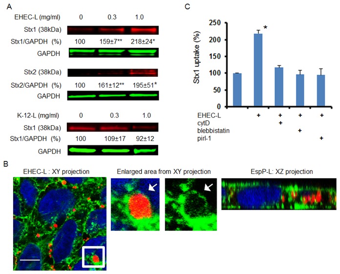
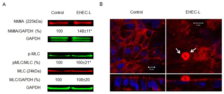
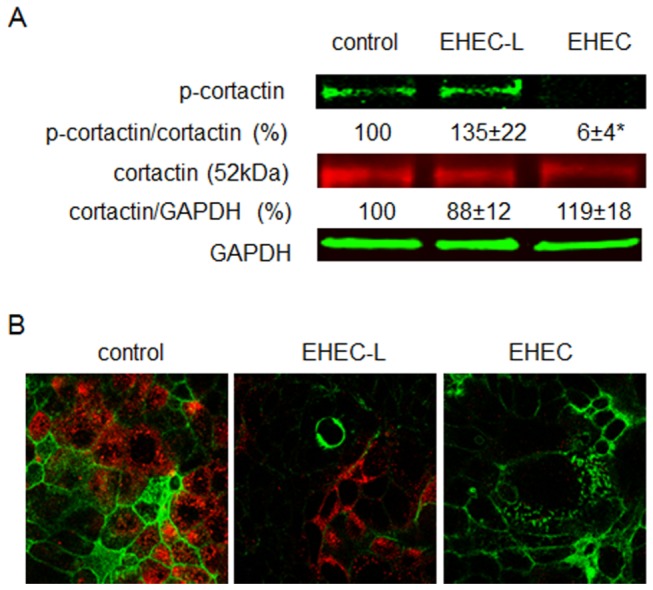
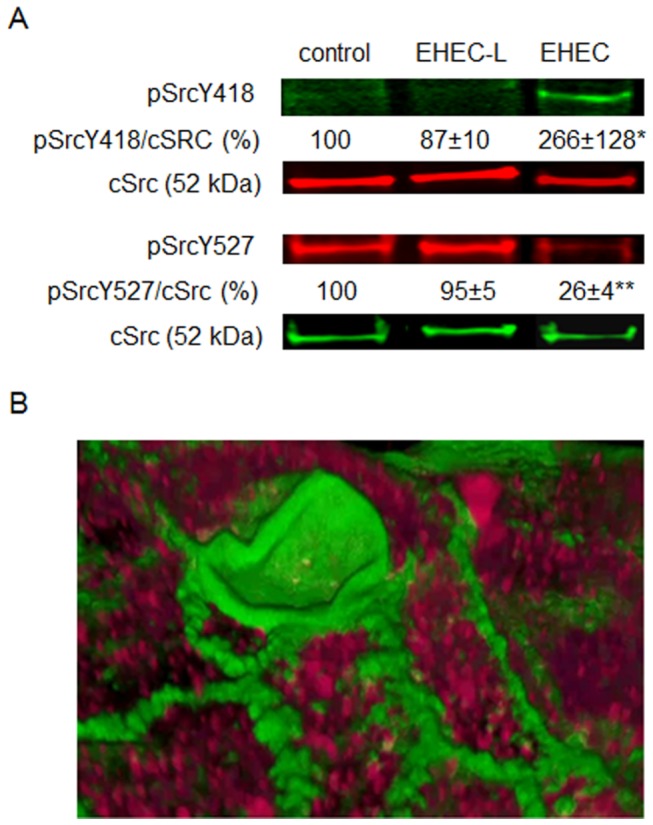
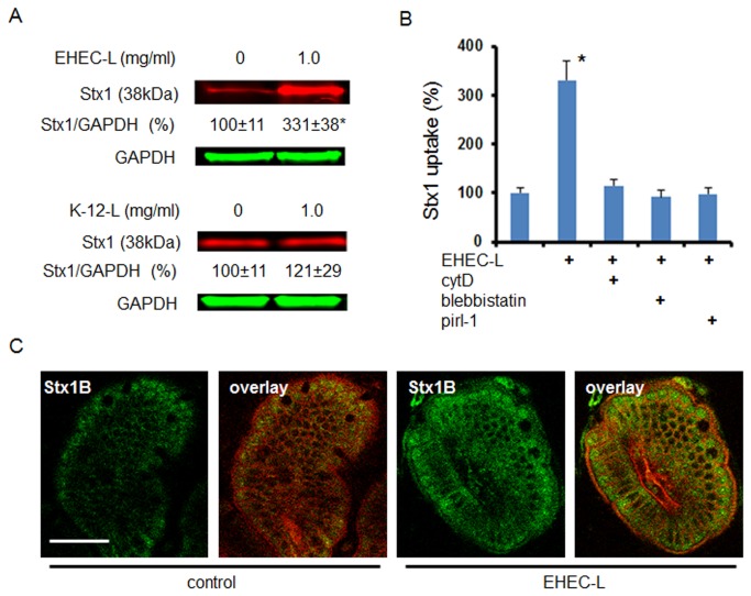
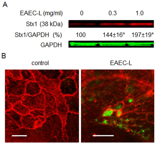
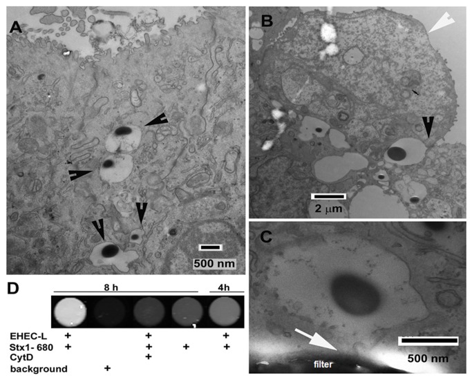
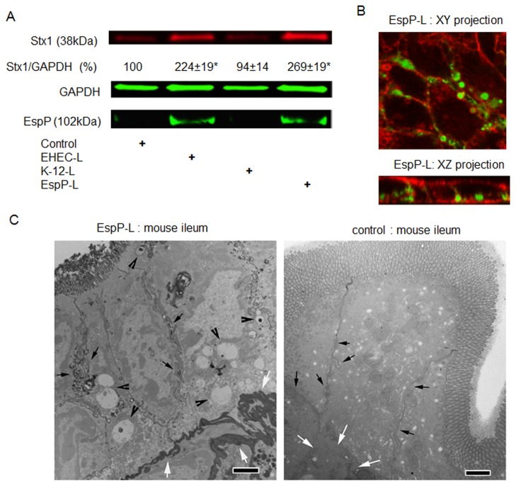
Life-threatening intestinal and systemic effects of the Shiga toxins produced by enterohemorrhagic Escherichia coli (EHEC) require toxin uptake and transcytosis across intestinal epithelial cells. We have recently demonstrated that EHEC infection of intestinal epithelial cells stimulates toxin macropinocytosis, an actin-dependent endocytic pathway. Host actin rearrangement necessary for EHEC attachment to enterocytes is mediated by the type 3 secretion system which functions as a molecular syringe to translocate bacterial effector proteins directly into host cells. Actin-dependent EHEC attachment also requires the outer membrane protein intimin, a major EHEC adhesin. Here, we investigate the role of type 3 secretion in actin turnover occurring during toxin macropinocytosis. Toxin macropinocytosis is independent of EHEC type 3 secretion and intimin attachment. EHEC soluble factors are sufficient to stimulate macropinocytosis and deliver toxin into enterocytes in vitro and in vivo; intact bacteria are not required. Intimin-negative enteroaggregative Escherichia coli (EAEC) O104:H4 robustly stimulate Shiga toxin macropinocytosis into intestinal epithelial cells. The apical macropinosomes formed in intestinal epithelial cells move through the cells and release their cargo at these cells' basolateral sides. Further analysis of EHEC secreted proteins shows that a serine protease EspP alone is able to stimulate host actin remodeling and toxin macropinocytosis. The observation that soluble factors, possibly serine proteases including EspP, from each of two genetically distinct toxin-producing strains, can stimulate Shiga toxin macropinocytosis and transcellular transcytosis alters current ideas concerning mechanisms whereby Shiga toxin interacts with human enterocytes. Mechanisms important for this macropinocytic pathway could suggest new potential therapeutic targets for Shiga toxin-induced disease.


Conflict of interest statement

Competing Interests: The authors have declared that no competing interests exist.

Figures

Figure 1
Figure 1. EHEC-L stimulates Stx1 and Stx2 uptake in T84 cells, while lysate from E. coli K12 strain does not.
(A) Representative immunoblots (IB) and quantitative representations of IB data show that increasing concentrations of EHEC-L significantly increase Stx1 and Stx2 uptake in T84 cells compared to untreated cells and cells treated with increasing concentrations of K12-L (n ≥ 6 monolayers per each experimental condition from 3 independent experiments; * - significant compared to the control (p < 0.05); ** - significant compared to control (p < 0.001). (B) Representative XY and XZ confocal optical section through T84 cells incubated for 4 h with EHEC-L (1 mg/mL) and Stx2 (0.5 µg/mL) show that EHEC reorganized actin in T84 cells and triggered the formation of actin coated macropinosomes of different sizes and many of the F-actin vesicle carry Stx2. Enlarged area from XY section (white box) shows that Stx1B is contained inside the F-actin-coated macropinosome. F-actin - green by phalloidin-AlexaFluor 488; Stx2 - red by AlexaFluor 568; nuclei – blue by Hoechst, bar -10 µm. (C) EHEC-L-induced Stx1 uptake was reduced to the control level in the presence of inhibitors of MPC including cytD, blebbistatin or pirl-1 (* - significant compared to control, p = 0.012; n ≥ 6 monolayers per each experimental condition from 3 independent experiments).
Figure 2
Figure 2. NMIIA and MLC are involved in EHEC-L-induced Stx MPC.
(A) Representative IB and quantitative data show that EHEC-L-induced MPC is accompanied by a significant increase in the relative amount of NMIIA and increase in pMLC (* - significant compared to the control (p < 0.01); n ≥ 4 monolayers per each experimental condition from 3 independent experiments. (B) Representative XY confocal optical sections through the apical region of T84 cells and corresponding XZ projections show the difference in MLC (red) distribution between control and EHEC-L-treated monolayers. White arrows indicate that upon EHEC-L treatment the MLC is concentrated in an apical macropinocytic bleb. In both panels: MLC-red by AlexaFluor568; nuclei – blue by Hoechst. Analysis of 23 apical F-actin blebs in EHEC-L treated cells from 2 independent experiments show that all 23 counted apical blebs were MLC-positive.
Figure 3
Figure 3
Cortactin is not involved in EHEC-L-induced MPC. (A) Representative IB and quantitative data show that treatment of T84 cells with EHEC-L does not affect the phosphorylation of cortactin (p-cortactin) in contrast to EHEC infection, which almost completely dephosphorylates cortactin (* p = 0.0006); n = 6 monolayers from 3 independent experiments). (B) Representative XY confocal optical sections through the apical region of T84 cells show that p-cortactin (red) is absent from the apical macropinocytic blebs detected by F-actin (green), but is present in surrounding cells not involved in MPC similar to that in control conditions. Analysis of 27 apical macropinocytic blebs in EHEC-L treated cells from 3 independent preparations showed no presence of p-cortactin in F-actin blebs. Also, p-cortactin is virtually absent from EHEC infected T84 monolayers. In both panels: p-cortactin – red by AlexaFluor568; F-actin – green by phalloidin-AlexaFluor488; nuclei – blue by Hoechst.
Figure 4
Figure 4
Src activation by EHEC infection is not involved in EHEC-stimulated MPC. (A) Representative IB and quantitative data show that treatment of T84 cells with EHEC-L does not activate Src (pSrcY418) in contrast to EHEC infection, which significantly increases the relative amount of pSrcY418 (* p = 0.044); n = 20 monolayers from10 independent experiments) and significantly decreases the relative amount of inactive pSrcY527 (** p = 0.0001, n = 20 monolayers from 10 independent experiments), while the relative amount of cSrc remains constant. (B) Representative 3D reconstruction of confocal optical sections through the apical region of T84 cells infected with EHEC strain EDL933 show that active pSrcY418 (red) is absent from the macropinocytic blebs detected by F-actin (green), but is present all though the cells. Analysis of 18 apical macropinocytic blebs in EDL933-infected cells from 2 independent preparations showed no presence of pSrcY418 in F-actin blebs. In panel B: pSrcY418 – red by AlexaFluor568; F-actin - green by phalloidin-AlexaFluor488.
Figure 5
Figure 5
EHEC-L stimulate Stx1 MPC in mouse ileum. (A) Representative IB and quantitative representations of data show that EHEC-L significantly increases Stx1 uptake in mouse enterocytes compared to tissue treated with K-12-L (n ≥ 6 animals per each experimental condition; * - significant compared to the control (p = 0.03)). (B) EHEC-L-induced Stx1 uptake in mouse intestine was reduced to the control level in the presence of inhibitors of MPC including cytD (n = 3 mice), blebbistatin (n = 4 mice) or pirl-1 (n = 4 mice). (C) Representative multiphoton optical section either through control sample of ileal tissue exposed to Stx1 only or tissue treated with EHEC-L plus Stx1 shows substantial increase in Stx1 fluorescence inside the enterocytes. In both panels: plasma membranes – red by tdTomato, Stx1-488 – green; bar -50 µm.
Figure 6
Figure 6
EAEC-L stimulates Stx1 uptake in T84 cells by stimulation of MPC. (A) Representative IB and quantitative representations of IB data show that increasing concentrations of EAEC-L significantly increased Stx1 uptake in T84 cells compared to untreated cells (n ≥ 3 monolayers per each experimental condition; * - significant compared to the control (p < 0.05)). (B) Representative XY optical sections through either control or EAEC-L-treated T84 cells additionally incubated with Stx1B-488 for 4 h show EHEC-L induced actin remodeling with formation of F-actin coated macropinosomes (spherical or irregularly shaped). Numerous macropinosomes carry the Stx1B-488 (green). F-actin - red by phalloidin -Alexa Fluor 568; bars -5 µm.
Figure 7
Figure 7
EHEC-L induced MPC leads to the transcellular transcytosis of the apical cargo. (A) Representative TEM image of T84 cells treated apically for 4h with a mixture of EHEC-L and 1 mg/mL HRP. EHEC-L causes the formation of macropinosomes filled with HRP (black arrowheads). (B) Representative TEM image depictures the process of a formation of HRP-bearing macropinosomes (black arrowhead). The apical EHEC-L induced bleb (white arrowhead) upon retraction back into the cell [19] and closure forms a new HRP-containing macropinosome. (C) Representative TEM image shows that the HRP-bearing macropinosome is reaching the basolateral side of filter-grown T84 cells (white arrow) and makes contact with the basal membrane. (D) Representative image obtained from fluorescence plate reader shows that EHEC-L stimulates Stx1 transcytosis in a time-dependent manner. This transcytosis is significantly inhibited by cytD (Table 2).
Figure 8
Figure 8
Serine protease EspP is sufficient to stimulate Stx1 MPC in T84 cells. (A) Representative IB and quantitative representations of IB data show that EspP expression by bacteria is sufficient to significantly increase Stx1 uptake in T84 cells compared to untreated cells or cells treated with lysates from K-12 bacteria that do not express EspP (n ≥ 3 monolayers per experimental condition; * - significant compared to the control (p < 0.05)). (B) Representative confocal optical sections through T84 cells incubated for 4 h in the presence of EspP-L (1 mg/mL) and B-subunit of Stx (Stx1B; 0.5 µg/mL) show that EspP reorganized actin in T84 cells and triggered the formation of actin coated macropinosomes that filled with Stx1B. F-actin - red by phalloidin-AlexaFluor568; Stx1B - green by AlexaFluor 488. (C) Representative TEM images of mouse ileal tissue treated from the luminal side with either a mixture of EspP-L and 2 mg/mL HRP or with mixture of K-12-L and 2 mg/mL HRP (control), bar -2 µm. EspP-L caused the formation of macropinosomes (black arrowheads) often containing HRP (black vesicles inside the macropinosomes). The macropinosomes are completely absent from control tissue. Importantly, EspP-L treatment caused the HRP accumulation in lamina propria (white arrows), which indicates the HRP transepithelial delivery. In contrast, HRP was absent from lamina propria (white arrows) in control tissue. Macropinosomes were often concentrated close to the lateral membranes (small black arrows) in ileal tissue, similar to observations in T84 cells, bars -2 µm.

Similar articles

Cited by

References

    1. Serna A 4th, Boedeker EC (2008) Pathogenesis and treatment of Shiga toxin-producing Escherichia coli infections. Curr Opin Gastroenterol 24: 38-47. doi:10.1097/MOG.0b013e3282f2dfb8. PubMed: 18043231. - DOI - PubMed
    1. Melton-Celsa A, Mohawk K, Teel L, O’Brien A (2012) Pathogenesis of Shiga-toxin producing escherichia coli. Curr Top Microbiol Immunol 357: 67-103. PubMed: 21915773. - PubMed
    1. Trachtman H, Austin C, Lewinski M, Stahl RA (2012) R e n a l a n d n e u r o l o g i c a l i n v o l v e m e n t in t y p i c a l S h i g a t o x i n - a s s o c i a t e d H U S . Nat Rev Nephrol 8(11):658-69 - PubMed
    1. Tarr PI, Gordon CA, Chandler WL (2005) Shiga-toxin-producing Escherichia coli and haemolytic uraemic syndrome. Lancet 365: 1073-1086. doi:10.1016/S0140-6736(05)74232-X. PubMed: 15781103. - DOI - PubMed
    1. Fuller CA, Pellino CA, Flagler MJ, Strasser JE, Weiss AA (2011) Shiga toxin subtypes display dramatic differences in potency. Infect Immun 79(3): 1329-1337. doi:10.1128/IAI.01182-10. PubMed: 21199911. - DOI - PMC - PubMed

Publication types