. 2013 Jul 9;8(7):e69330.
doi: 10.1371/journal.pone.0069330. Print 2013.

Fgf21 impairs adipocyte insulin sensitivity in mice fed a low-carbohydrate, high-fat ketogenic diet

Affiliations

Fgf21 impairs adipocyte insulin sensitivity in mice fed a low-carbohydrate, high-fat ketogenic diet

Yusuke Murata et al. PLoS One. .

Abstract

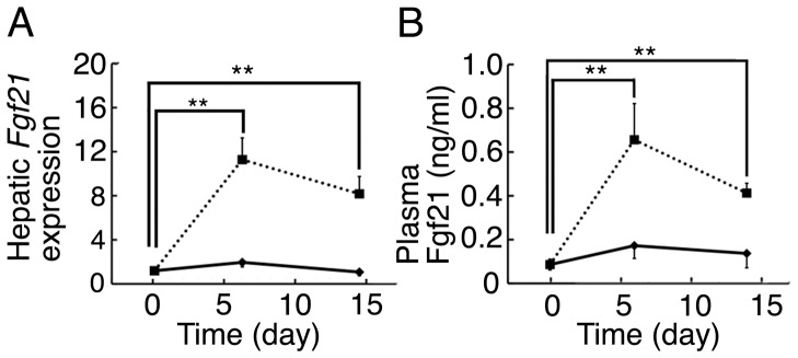
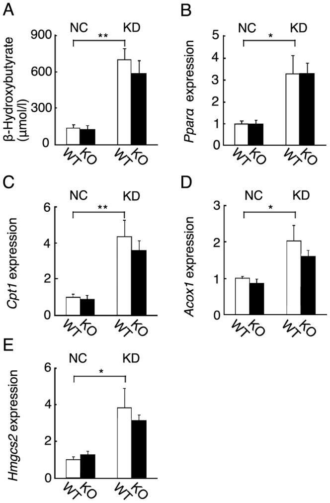
Background: A low-carbohydrate, high-fat ketogenic diet (KD) induces hepatic ketogenesis and is believed to affect energy metabolism in mice. As hepatic Fgf21 expression was markedly induced in mice fed KD, we examined the effects of KD feeding on metabolism and the roles of Fgf21 in metabolism in mice fed KD using Fgf21 knockout mice.

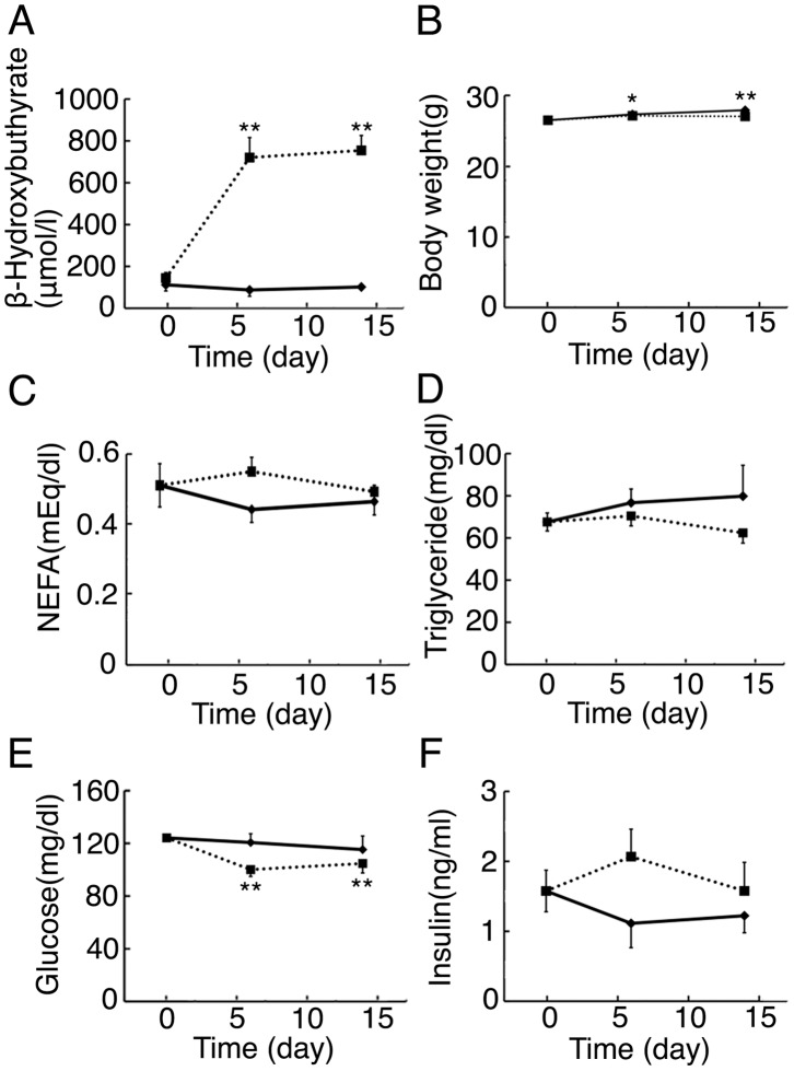
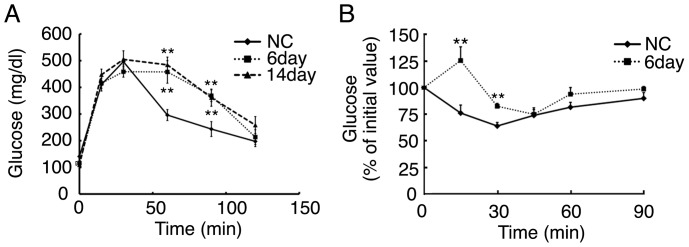
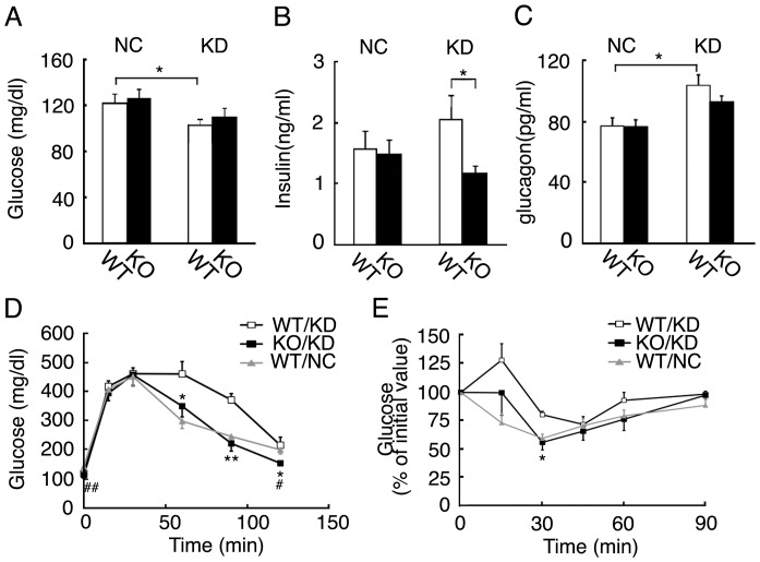
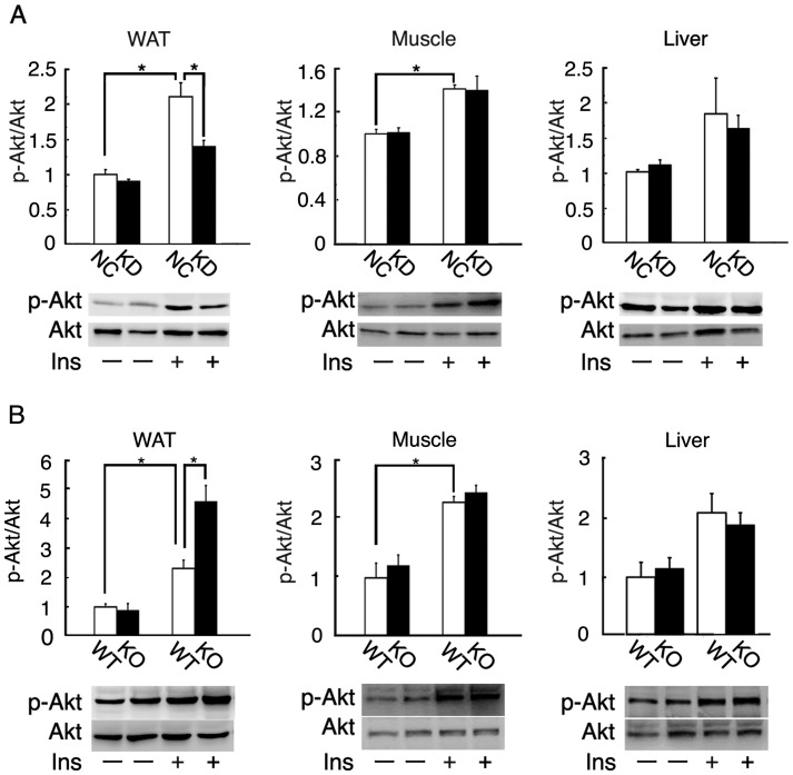
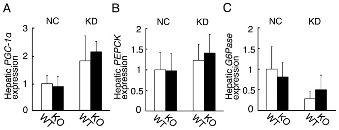
Methodology/principal findings: We examined C57BL/6 mice fed KD for 6 or 14 days. Blood β-hydroxybutyrate levels were greatly increased at 6 days, indicating that hepatic ketogenesis was induced effectively by KD feeding for 6 days. KD feeding for 6 and 14 days impaired glucose tolerance and insulin sensitivity, although it did not affect body weight, blood NEFA, and triglyceride levels. Hepatic Fgf21 expression and blood Fgf21 levels were markedly increased in mice fed KD for 6 days. Blood β-hydroxybutyrate levels in the knockout mice fed KD for 6 days were comparable to those in wild-type mice fed KD, indicating that Fgf21 is not required for ketogenesis. However, the impaired glucose tolerance and insulin sensitivity caused by KD feeding were improved in the knockout mice. Insulin-stimulated Akt phosphorylation was significantly decreased in the white adipose tissue in wild-type mice fed KD compared with those fed normal chow, but not in the muscle and liver. Its phosphorylation in the white adipose tissue was significantly increased in the knockout mice fed KD compared with wild-type mice fed KD. In contrast, hepatic gluconeogenic gene expression in Fgf21 knockout mice fed KD was comparable to those in the wild-type mice fed KD.

Conclusions/significance: The present findings indicate that KD feeding impairs insulin sensitivity in mice due to insulin resistance in white adipose tissue. In addition, our findings indicate that Fgf21 induced to express by KD is a negative regulator of adipocyte insulin sensitivity in adaptation to a low-carbohydrate malnutritional state.


Conflict of interest statement

Competing Interests: The authors have declared that no competing interests exist.

Figures

Figure 1
Figure 1. Body weights and blood parameters in mice fed KD.
(A) Blood β-hydroxybutyrate levels in 3-month-old mice fed KD for 6 days or 14 days, or NC (n = 3–8 per group; **, P<0.01 vs. day 0). (B) Body weights in 3-month-old mice fed KD for 6 days or 14 days, or NC (n = 5–28per group; *, P<0.05; **, P<0.01 vs. day 0). (C–F) Blood NEFA, triglyceride, glucose, and insulin levels in 3-month-old mice fed KD for 6 days or 14 days, or NC (n = 4–12 per group; **, P<0.01 vs. day 0).
Figure 2
Figure 2. Glucose tolerance and insulin tolerance tests of mice fed KD.
(A) A glucose tolerance test (GTT) of 3-month-old mice fed KD for 6 days or 14 days, or NC (n = 8–11 per group; **, P<0.01 vs. mice fed NC at the same timepoint). (B) An insulin tolerance test of mice fed KD for 6 days or 14 days, or NC (n = 4–7 per group; **, P<0.01 vs. mice fed NC at the same timepoint).
Figure 3
Figure 3. Hepatic Fgf21 expression and blood Fgf21 levels of mice fed KD.
(A) Hepatic Fgf21 expression of 3-month-old mice fed KD for 6 days or 14 days, or NC determined by RT-qPCR (n = 4–8 per group; **, P<0.01 vs. day 0). (B) Blood Fgf21 levels of 3-month-old mice fed KD for 6 days or 14 days, or NC determined by ELISA (n = 5–7 per group; **, P<0.01 vs. day 0).
Figure 4
Figure 4. Body and tissue weights of wild-type and Fgf21 knockout mice fed NC or KD.
(A) Body weights of 3-month-old wild-type (WT) and Fgf21 knockout (KO) mice fed NC or KD for 6 days (n = 7–8 mice per group). (B–E) The heart, kidney, liver, and subcutaneous WAT weights of 3-month-old wild-type (WT) and Fgf21 knockout (KO) mice fed NC or KD for 6 days (n = 7–8 mice per group).
Figure 5
Figure 5. Hepatic ketogenesis of wild-type and Fgf21 knockout mice.
(A) Blood β-hydroxybutyrate levels in 3-month-old wild-type (WT) and Fgf21 knockout (KO) mice fed NC or KD for 6 days. (n = 12–14 mice per group; **, P<0.01). (B–E) Hepatic expression of genes involved in ketogenesis including Ppara, Cpt1, Acox1, and Hmgcs2 in wild-type (WT) and Fgf21 knockout (KO) mice fed NC or KD for 6 days. (n = 7–9 mice per group; *, P<0.05; **, P<0.01).
Figure 6
Figure 6. Glucose tolerance and insulin tolerance tests of wild-type and Fgf21 knockout mice.
(A–C) Blood glucose, insulin, and glucagon levels of 3-month-old wild-type (WT) and Fgf21 knockout (KO) mice fed NC or KD for 6 days (n = 8–12 mice per group; *, P<0.05). (D) A glucose tolerance test (GTT) of 3-month-old wild-type (WT) and Fgf21 knockout (KO) mice fed KD for 6 days (n = 6–10 per group; *, P<0.05; **, P<0.01 vs. wild-type mice fed KD at the same timepoint, #, P<0.05; P<0.01 vs. wild-type mice fed NC at the same timepoint). (E) An insulin tolerance test of wild-type (WT) mice fed NC or KD and Fgf21 knockout (KO) mice fed KD for 6 days (n = 4–7 per group; *, P<0.05 vs. wild-type mice fed KD at the same timepoint).
Figure 7
Figure 7. Insulin-stimulated Akt phosphorylation of wild-type and Fgf21 knockout mice.
(A) Basal or insulin-stimulated Akt phosphorylation in the subcutaneous white adipose tissue, gastrocnemius muscle, and liver of 3-month-old wild-type mice fed NC or KD for 6 days (n = 7 per group; *, P<0.05). (B) Basal or insulin-stimulated Akt phosphorylation in the subcutaneous white adipose tissue, gastrocnemius muscle, and liver of 3-month-old wild-type (WT) and Fgf21 knockout (KO) mice fed KD for 6 days (n = 7 per group; *, P<0.05). Akt and the phosphorylated form were detected by Western blotting with specific antibodies.
Figure 8
Figure 8. Hepatic expression of key genes involved in gluconeogenesis in wild-type and Fgf21 knockout mice fed NC or KD.
Hepatic expression of PGC-1α, PEPCK, and G6Pase in wild-type (WT) and Fgf21 knockout (KO) mice fed NC or KD for 6 days. (n = 6–8 mice per group).

Similar articles

Cited by

References

    1. Itoh N, Ornitz DM (2008) Functional evolutionary history of the mouse Fgf gene familiy. Dev. Dyn. 237: 18–27. - PubMed
    1. Itoh N, Ornitz DM (2011) Fibroblast growth factors: from molecular evolution to roles in development, metabolism and disease. J. Biochem. 149: 121–130. - PMC - PubMed
    1. Nishimura T, Nakatake Y, Konishi M, Itoh N (2000) Identification of a novel FGF, FGF-21, preferentially expressed in the liver. Biochim. Biophys. Acta. 1492: 203–206. - PubMed
    1. Kharitonenkov A, Shiyanova TL, Koester A, Ford AM, Micanovic R, et al. (2005) FGF21 as a novel metabolic regulator. J. Clin. Invest. 115: 1627–1635. - PMC - PubMed
    1. Kharitonenkov A, Larsen P (2011) FGF21 reloaded: challenges of a rapidly growing field. Trends Endocrinol. Metab. 22: 81–86. - PubMed

Publication types

MeSH terms

LinkOut - more resources