. 2013 Jul 11;8(7):e69677.
doi: 10.1371/journal.pone.0069677. Print 2013.

4-1BB signaling activates the t cell factor 1 effector/β-catenin pathway with delayed kinetics via ERK signaling and delayed PI3K/AKT activation to promote the proliferation of CD8+ T Cells

Affiliations

4-1BB signaling activates the t cell factor 1 effector/β-catenin pathway with delayed kinetics via ERK signaling and delayed PI3K/AKT activation to promote the proliferation of CD8+ T Cells

Do Y Lee et al. PLoS One. .

Abstract

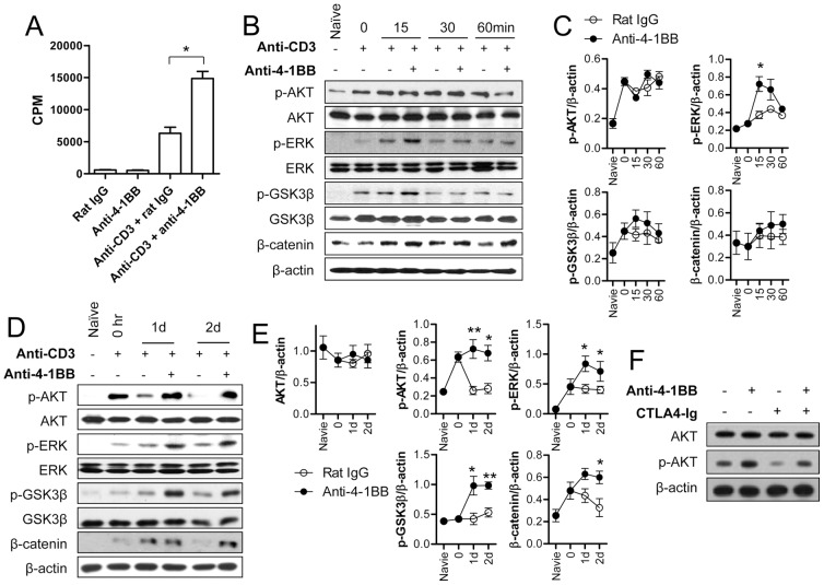
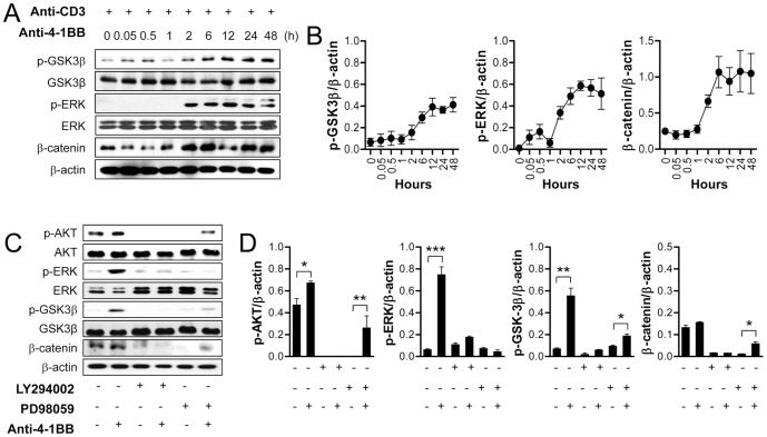
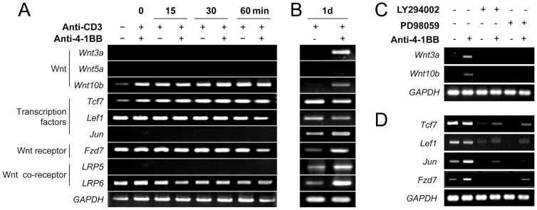
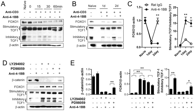
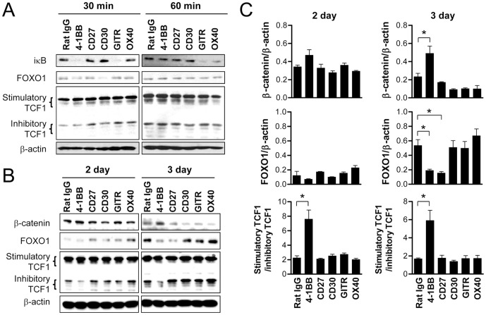
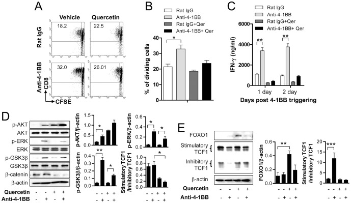
4-1BB (CD137), an inducible costimulatory molecule, strongly enhances the proliferation and effector function of CD8(+) T cells. Since the serine/threonine kinase, glycogen synthase kinase-3 (GSK-3), is involved in a variety of signaling pathways of cellular proliferation, migration, immune responses, and apoptosis, we examined whether 4-1BB signaling activates GSK-3/β-catenin signaling and downstream transcription factors to enhance the proliferation of CD8(+) T cells. 4-1BB signaling induces rapid activation of ERK and IκB degradation, and shows delayed activation of AKT at 24 h post 4-1BB stimulation on anti-CD3 activated T cells. ERK and AKT signals were required for sustained β-catenin levels by inactivating GSK-3, which was also observed with delayed kinetics after 4-1BB stimulation. As a transcriptional partner of β-catenin, 4-1BB signaling decreased levels of FOXO1 and increased levels of stimulatory TCF1 in CD8(+) T cells at 2-3 days but not at early time points after 4-1BB engagement. The enhanced proliferation of CD8(+) T cells due to 4-1BB signaling was completely abolished by treatment with the TCF1/β-catenin inhibitor quercetin. These results show that 4-1BB signaling enhances the proliferation of activated CD8(+) T cells by activating the TCF1/β-catenin axis via the PI3K/AKT/ERK pathway. As effects of 4-1BB on AKT, FOXO1, β-catenin and GSK-3β showed delayed kinetics it is likely that an intervening molecule induced by 4-1BB and ERK signaling in activated T cells is responsible for these effects. These effects were observed on CD8(+) but not on CD4(+) T cells. Moreover, 4-1BB appeared to be unique among several TNFRs tested in inducing increase in stimulatory over inhibitory TCF-1.


Conflict of interest statement

Competing Interests: The authors have declared that no competing interests exist.

Figures

Figure 1
Figure 1. 4-1BB sustains anti-CD3 induced signals to induce GSK-3/β-catenin signaling with delayed kinetics.
The purified CD8+ T cells from C57BL/6 mice were activated with 0.1 µg/ml of anti-CD3 for 16 h. (A) CD8+ T cells cultured with or without anti-CD3 for 16 h were further incubated with 5 µg/ml of anti-4-1BB or rat IgG for another 48 h, and cell proliferation was assessed by [3H]-thymidine incorporation. (B–C) The activated CD8+ T cells were incubated with 5 µg/ml of anti-4-1BB or rat IgG for 0, 15, 30, or 60 min. Western blot analysis was performed with p-AKT, p-ERK, p-GSK-3β, β-catenin, and β-actin antibodies (B) and the relative expression levels of p-AKT, p-ERK, p-GSK-3β, and β-catenin were determined (C). (D–E) The activated CD8+ T cells were stimulated with 5 µg/ml of anti-4-1BB for 0, 1, or 2 days. Western blotting was performed (D) and the relative expression levels of each protein was determined (E). (F) The activated CD8+ T cells were treated with 5 µg/ml anti-4-1BB or rat IgG following 1 h incubation with 10 µg/ml CTLA-4Ig. After 24 h incubation, CD8+ T cells were harvested, lysed and subjected to Western blot analysis using AKT, p-AKT, and β-catenin antibodies. Naïve indicates CD8+ T cells that were cultured in complete medium for 16 h without anti-CD3. Results in C and E are mean ±SD of three independent experiments and representative data of two independent experiments are shown in F (*p<0.05; **p<0.01).
Figure 2
Figure 2. GSK-3/β-catenin signaling downstream of anti-CD3 plus anti-4-1BB is dependent on PI3K-ERK pathway.
(A–B) CD8+ T cells activated by anti-CD3 for 16 h were stimulated with 5 µg/ml of anti-4-1BB for the indicated times. Western blotting was performed with p-GSK-3β, p-ERK, β-catenin, and β-actin antibodies (A), and the relative expression levels were calculated by dividing the densitometry value of each protein by the value of β-actin (B). (C–D) The activated CD8+ T cells were pre-incubated with or without 10 µM LY294002 or 10 µM PD98509 and then stimulated with 5 µg/ml of anti-4-1BB or rat IgG for 1 day. Western blotting was performed with p-AKT, p-GSK-3β, p-ERK, and β-catenin antibodies (C) and the relative expression levels of each protein was determined (D). Results in B and D are mean ±SD of three separate experiments (*p<0.05; **p<0.01; ***p<0.001).
Figure 3
Figure 3. 4-1BB triggering increases levels of Wnt ligands and their receptor.
(A–B) CD8+ T lymphocytes were activated with anti-CD3 and then stimulated with anti-4-1BB or rat IgG for the indicated times. Total RNA was extracted from the CD8+ T cells and transcripts of Wnts, Tcf7, Lef, Fzd, Jun, and Wnt co-receptor LRP5/6 were measured by RT-PCR. GAPDH was used as an internal control. (C–D) The activated CD8+ T cells were pre-incubated with or without 10 µM LY294002 or 10 µM PD98059 for 1 h and then stimulated with 5 µg/ml anti-4-1BB or rat IgG for 24 h. Total RNAs were extracted and RT-PCR was performed with specific primer sets. Representative data from at least three independent experiments are shown.
Figure 4
Figure 4. 4-1BB signaling induces the delayed activation of β-catenin/TCF1 pathway under the control of PI3K-ERK pathway.
(A–C) Anti-CD3-activated CD8+ T cells were stimulated with 5 µg/ml anti-4-1BB or rat IgG for the indicated times. Western blotting was performed with antibodies against β-catenin, TCF1, FOXO1 and β-actin. (A) Expression levels of FOXO1, TCF1 and β-catenin 0, 15, 30, or 60 min after 4-1BB triggering. (B) Expression levels of FOXO1, TCF1 and β-catenin 1 or 2 days after 4-1BB triggering. (C) The relative expression levels of FOXO1 and TCF1 on days 1 and 2 were calculated. (D–E) Activated CD8+ T cells were pre-incubated with or without 10 µM LY294002 or 10 µM PD98509 for 1 h and then stimulated with anti-4-1BB or rat IgG for 24 h. Western blotting was performed with β-catenin, FOXO1, TCF1, and β-actin antibodies (D), and the relative expression levels of β-catenin, FOXO1 and TCF1 were calculated (E). Naïve indicates CD8+ T cells that were cultured in complete medium for 16 h without anti-CD3. Results in C and E are mean ±SD of three separate experiments (*p<0.05; **p<0.01).
Figure 5
Figure 5. 4-1BB is unique among TNFRSF members in activating the β-catenin/TCF1 pathway in CD8+ T cells.
(A) CD8+ T cells activated by anti-CD3 for 16 h were stimulated with 5 µg/ml of anti-4-1BB, anti-CD27, and anti-CD30, or 10 µg/ml anti-GITR and anti-OX40 for 30 or 60 min. Western blot analysis was performed using IκB, FOXO1, TCF1, and β-actin antibodies. (B–C) Freshly isolated CD8+ T cells were activated with 0.1 µg/ml of anti-CD3 and simultaneously treated with 5 µg/ml of anti-4-1BB, anti-CD27, and anti-CD30, or 10 µg/ml anti-GITR and anti-OX40 for 2 or 3 days. Western blotting was performed with antibodies specific for β-catenin, TCF1 and FOXO1 and β-actin (B). The relative expression levels of β-catenin, FOXO1 and TCF1 were calculated (C). Results are mean ±SD of two separate experiments (*p<0.05).
Figure 6
Figure 6. 4-1BB signaling activates the β-catenin/TCF1 pathway in CD8+ T cells but not in CD4+ T cells.
(A–B) CD8+ T cells were isolated from WT and 4-1BB−/− C57BL/6 mice, activated with anti-CD3 for 16 h, and incubated with anti-4-1BB for 1 or 2 days. Western blotting was performed with antibodies specific for β-catenin, TCF1, FOXO1 and β-actin (A). The relative expression levels of β-catenin, FOXO1 and TCF1 were calculated (B). (C) Purified CD4+ and CD8+ T cells were activated with the indicated dose of anti-CD3 mAb for 24 h. The activated T cells were stained with FITC-conjugated anti-CD4 or anti-CD8 mAb along with PE-conjugated anti-4-1BB mAb (eBioscience) and subsequently analyzed by FACSCalibur (BD Bioscience). (D–G) CD8+ and CD4+ T cells were activated for 16 h and 36 h, respectively in the presence of 0.1 µg/ml anti-CD3, and then stimulated with 5 µg/ml 4-1BB for the indicated times. (D) FACS analysis of CD4+ and CD8+ T cells. (E) Expression levels of FOXO1, TCF1 and β-catenin in activated CD8+ T and CD4+ T cells 0, 15, or 30 min after 4-1BB triggering. (F) Expression levels of FOXO1, TCF1 and β-catenin in activated CD8+ T and CD4+ T cells 1 or 2 days after 4-1BB triggering. (G) The relative expression levels of β-catenin, FOXO1 and TCF1 were calculated from E. Naïve indicates CD8+ T cells that were cultured in complete medium for 16 h without anti-CD3. Results are mean ±SD of three separate experiments (*p<0.05).
Figure 7
Figure 7. 4-1BB signaling enhances the proliferation of CD8+ T cells and their IFN-γ production through β-catenin/TCF1 interaction.
(A–B) Purified CD8+ T cells labeled with CFSE were incubated with 0.1 µg/ml of anti-CD3 for 16 h. The activated CD8+ T cells were pre-incubated with or without 25 µM quercetin for 1 h and then stimulated with anti-4-1BB or rat IgG for 48 h. Cell proliferation was assessed by the CFSE dilution method. Live cells were gated, and plotted as CFSE vs. CD8 (A), and proportions of the dividing cells were determined (n = 3) (B). (C) Activated CD8+ T cells were pre-incubated with or without 25 µM quercetin for 1 h and then stimulated with anti-4-1BB or rat IgG for 24 h or 48 h. The IFN-γ concentration in cell supernatants was determined using a BD™ Cytometric Bead Array kit (BD Bioscience). (D, E) Activated CD8+ T cells were pre-incubated with or without 25 µM quercetin for 1 h and then stimulated with anti-4-1BB or rat IgG for 24 h. Western blotting was performed with specific antibodies and the relative expression levels of p-AKT, p-ERK, p-GSK-3β and β-catenin were calculated. Results are mean ±SD of three separate experiments (*p<0.05; **p<0.01).

References

    1. Hurtado JC, Kim YJ, Kwon BS (1997) Signals through 4-1BB are costimulatory to previously activated splenic T cells and inhibit activation-induced cell death. J Immunol 158: 2600–2609. - PubMed
    1. Halstead ES, Mueller YM, Altman JD, Katsikis PD (2002) In vivo stimulation of CD137 broadens primary antiviral CD8+ T cell responses. Nat Immunol 3: 536–541. - PubMed
    1. Lee HW, Park SJ, Choi BK, Kim HH, Nam KO, et al. (2002) 4-1BB promotes the survival of CD8+ T lymphocytes by increasing expression of Bcl-xL and Bfl-1. J Immunol 169: 4882–4888. - PubMed
    1. Lee HW, Nam KO, Park SJ, Kwon BS (2003) 4-1BB enhances CD8+ T cell expansion by regulating cell cycle progression through changes in expression of cyclins D and E and cyclin-dependent kinase inhibitor p27kip1. Eur J Immunol 33: 2133–2141. - PubMed
    1. Vinay DS, Kwon BS (2012) Immunotherapy of cancer with 4-1BB. Mol Cancer Ther 11: 1062–1070. - PubMed

Publication types

MeSH terms

Substances