. 2013 Jun 15;2(1):263.
doi: 10.1186/2193-1801-2-263. Print 2013 Dec.

A simple set of validation steps identifies and removes false results in a sandwich enzyme-linked immunosorbent assay caused by anti-animal IgG antibodies in plasma from arthritis patients

Affiliations

A simple set of validation steps identifies and removes false results in a sandwich enzyme-linked immunosorbent assay caused by anti-animal IgG antibodies in plasma from arthritis patients

Tue W Kragstrup et al. Springerplus. .

Abstract

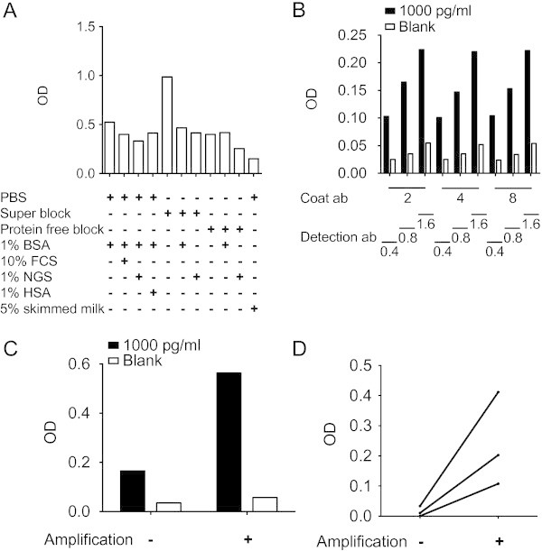
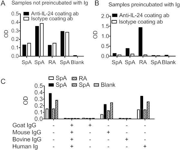
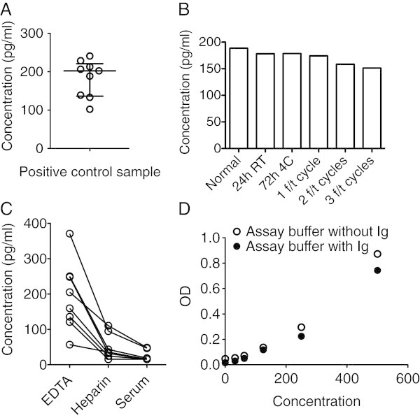
Rheumatoid arthritis (RA) and spondyloarthritis (SpA) are chronic diseases characterized by activation of the immune system and production of antibodies. Thus, rheumatoid factor, anti-animal IgG antibodies and heterophilic antibodies in plasma samples from arthritis patients can interfere with immunoassays such as sandwich enzyme-linked immunosorbent assay (ELISA) systems often used in arthritis research. However, standard methodologies on how to test for false results caused by these antibodies are lacking. The objective of this study was to design a simple set of steps to validate a sandwich ELISA before using it for measuring analytes in plasma from arthritis patients. An interleukin-24 (IL-24) sandwich ELISA system was prepared with a monoclonal mouse capture antibody and a polyclonal goat detection antibody and tested for interference by rheumatoid factor, anti-animal IgG antibodies and heterophilic antibodies. Plasma samples from 23 patients with RA and SpA were used. No differences were found between plasma samples measured in wells coated with anti-IL-24 specific antibody and in wells coated with isotype control antibody (false positive results), and recombinant human IL-24 was not recovered in spiked samples (false negative results). This interference was removed after preincubating the plasma samples from patients with arthritis with goat or bovine IgG, suggesting that anti-animal IgG antibodies found in the plasma of the arthritis patients caused the false results. Additional testing showed that the signal-to-noise ratio could be increased by titration of the capture and detection antibodies and by using the ELAST amplification system. Finally, the calculated concentration of IL-24 was increased in ethylenediaminetetraacetic acid (EDTA) plasma compared to heparin plasma and serum and decreased with repetitive freeze/thaw cycles of the samples illustrating how sample handling could additionally contribute to the variations reported by different laboratories in measurement of the same analyte. This study proposes a simple set of validation steps to evaluate and optimize a sandwich ELISA before using it for measuring analytes in plasma from arthritis patients. Anti-animal IgG antibodies are also present in healthy individuals, suggesting that validation of ELISA systems for measuring non-arthritis samples could also be improved by this simple set of validation steps.

Keywords: Anti-animal IgG antibodies; Arthritis; ELAST amplification system; Enzyme-linked immunosorbent assay; Heterophilic antibodies; Immunoassay; Interference; Multiplex; Rheumatoid factor.


Figures

Figure 1
Figure 1
Preparing the sandwich ELISA system. (A) Testing different blockers of non-specific binding sites in polystyrene wells. OD signal from wells with blank assay diluent after blocking the wells with different blocking reagents. (B) Titration of capture and detection antibodies. OD signal from wells with blank assay diluent (noise) and wells with 1000 pg/ml rh IL-24 in assay diluent (signal) using different concentrations of capture antibody and detection antibody. Numbers represent concentrations in μg/ml. (C) OD signal from wells with blank assay diluent (noise) and wells with 1000 pg/ml rh IL-24 in assay diluent (signal) without or with amplification. (D) OD signal from wells with plasma samples from three different arthritis patients without or with amplification.
Figure 2
Figure 2
Testing for false positive results. (A) False positive measurements. OD signal from four patient samples in wells coated with anti-IL-24 specific antibody or with isotype control antibody. (B) Identification and removal of false positive measurements. OD signal from the same four patient samples in wells coated with anti-IL-24 specific antibody or with isotype control antibody after preincubating samples with a combination of mouse, goat, bovine and human immunoglobulins. (C) Identification of species specificity of the anti-animal IgG antibodies in the patient samples. OD signal from the same four patient samples in wells coated with isotype antibody after preincubating samples with different immunoglobulins as indicated.
Figure 3
Figure 3
Testing for false negative results and other matrix effects. (A) False negative measurements. OD signal from unspiked and spiked plasma samples from two arthritis patients in wells coated with anti-IL-24 specific antibody. (B) Identification and removal of false negative measurements. OD signal from unspiked and spiked plasma samples from the same two arthritis patients in wells coated with anti-IL-24 specific antibody after preincubating samples with a combination of mouse, goat, bovine and human immunoglobulins. (C) Spike recovery assessment in three other arthritis patients. Calculated IL-24 concentrations from unspiked patient samples and patient samples spiked with 800 pg/ml rh IL-24 in wells coated with anti-IL-24 specific antibody after preincubating samples with immunoglobulins. (D) Linearity-of-dilution assessment in three other arthritis patients. Calculated IL-24 concentrations in samples preincubated with immunoglobulin and diluted 4/6, 2/6 and 1/6 in wells coated with anti-IL-24 specific antibody.
Figure 4
Figure 4
Additional testing of the ELISA system. (A) Testing the ELISA system for variability. Calculated IL-24 concentrations in the positive control sample analyzed in nine different experiments. (B) Testing the ELISA system for stability of the analyte. Calculated IL-24 concentrations in the positive control sample after storage under different conditions or after a variable number of freeze/thaw cycles (f/t). (C) Testing the ELISA system for effect of the sample anticoagulant used. Calculated IL-24 concentrations in eight paired plasma samples from patients with arthritis stabilized with either EDTA or heparin or serum samples. (D) Testing the ELISA system for effect on the standard curve of adding immunoglobulins to the assay diluent. OD signal from samples of rh IL-24 diluted in assay buffer without immunoglobulin and from samples of rh IL-24 diluted in assay buffer with immunoglobulin.

References

    1. Andersen DC, Koch C, Jensen CH, et al. High prevalence of human anti-bovine IgG antibodies as the major cause of false positive reactions in two-site immunoassays based on monoclonal antibodies. J Immunoassay Immunochemistry. 2004;25:17–30. doi: 10.1081/IAS-120027223. - DOI - PubMed
    1. Arnett FC, Edworthy SM, Bloch DA, et al. The American Rheumatism Association 1987 revised criteria for the classification of rheumatoid arthritis. Arthritis Rheum. 1988;31:315–324. doi: 10.1002/art.1780310302. - DOI - PubMed
    1. Ball J, Lawrence JS. Epidemiology of the sheep cell agglutination test. Ann Rheum Dis. 1961;20:235–243. doi: 10.1136/ard.20.3.235. - DOI - PMC - PubMed
    1. Bartels EM, Ribel-Madsen S. Methods. 2013. Cytokine measurements and possible interference from heterophilic antibodies - Problems and solutions experienced with rheumatoid factor. - PubMed
    1. Bartels EM, Falbe Wätjen I, Littrup Andersen E, et al. Rheumatoid factor and its interference with cytokine measurements: problems and solutions. Arthritis. 2011;2011:741071. doi: 10.1155/2011/741071. - DOI - PMC - PubMed