Review
. 2013 Jul 16:3:26.
doi: 10.3389/fcimb.2013.00026. eCollection 2013.

Interaction of the tick immune system with transmitted pathogens

Affiliations
Review

Interaction of the tick immune system with transmitted pathogens

Ondřej Hajdušek et al. Front Cell Infect Microbiol. .

Abstract

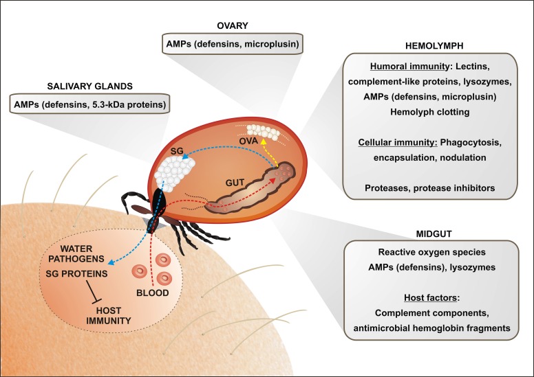
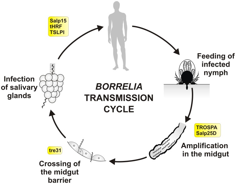
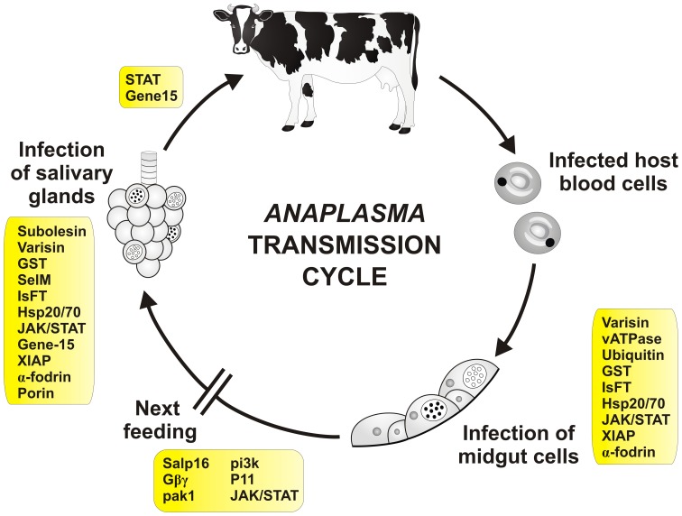
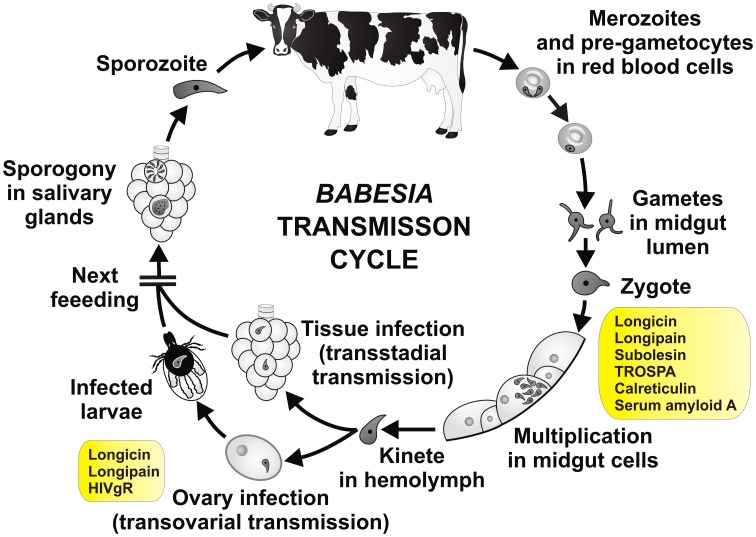
Ticks are hematophagous arachnids transmitting a wide variety of pathogens including viruses, bacteria, and protozoans to their vertebrate hosts. The tick vector competence has to be intimately linked to the ability of transmitted pathogens to evade tick defense mechanisms encountered on their route through the tick body comprising midgut, hemolymph, salivary glands or ovaries. Tick innate immunity is, like in other invertebrates, based on an orchestrated action of humoral and cellular immune responses. The direct antimicrobial defense in ticks is accomplished by a variety of small molecules such as defensins, lysozymes or by tick-specific antimicrobial compounds such as microplusin/hebraein or 5.3-kDa family proteins. Phagocytosis of the invading microbes by tick hemocytes is likely mediated by the primordial complement-like system composed of thioester-containing proteins, fibrinogen-related lectins and convertase-like factors. Moreover, an important role in survival of the ingested microbes seems to be played by host proteins and redox balance maintenance in the tick midgut. Here, we summarize recent knowledge about the major components of tick immune system and focus on their interaction with the relevant tick-transmitted pathogens, represented by spirochetes (Borrelia), rickettsiae (Anaplasma), and protozoans (Babesia). Availability of the tick genomic database and feasibility of functional genomics based on RNA interference greatly contribute to the understanding of molecular and cellular interplay at the tick-pathogen interface and may provide new targets for blocking the transmission of tick pathogens.

Keywords: Anaplasma; Babesia; Borrelia; antimicrobial peptides; innate immunity; phagocytosis; tick; tick-borne diseases.


Figures

Figure 1
Figure 1
An overview of the tick immune mechanisms and molecules constituting potential barriers for the pathogen transmission. The pathogen transmission is tightly linked with physiology of blood feeding and tick innate immunity. Ingested blood meal is accumulated in the midgut content (red arrow; only one caecum shown). Hemoglobin and other proteins are taken up by the tick midgut cells and digested intracellularly in the lysosome-like digestive vesicles (Sojka et al., 2013). Liberated amino acids and other compounds are transported to the peripheral tissues and ovaries, supplying mainly egg development (yellow arrow). Importantly, the blood meal is concentrated by reabsorption of excessive water, which is spitted back into the wound by the action of salivary glands (blue arrow). Tick saliva contains a great variety of anti-coagulant, immunomodulatory and anti-inflammatory molecules that facilitate pathogen acquisition and transmission. The ingested pathogens have to survive the period between detachment and subsequent feeding of the next tick developmental stage and overcome several obstacles on its route through the tick body. In the midgut, tick may utilize some of the host immune molecules (e.g., complement system) for its own defense against intestinal inhabitants. Hemoglobin fragments, derived from the host hemoglobin, are secreted into the midgut lumen and exert strong antimicrobial activity. Tick midgut tissue also expresses a variety of endogenous AMPs, which sustain the midgut microbes at a tolerable level. An important, but still poorly understood, role is most likely played by the maintenance of the redox homeostasis in the tick midgut. Pathogens intruding into the tick hemocoel can be phagocytosed by tick hemocytes or destroyed by effector molecules of the humoral defense system, comprising AMPs, components of the primordial complement system (thioester-containing proteins (TEPs), convertase-like factors and fibrinogen-related lectins (FREPs). Ticks probably possess a mechanism of hemolymph clotting, but genes/proteins putatively involved in the activation of prophenoloxidase cascade leading to melanization have not yet been identified in any tick species. Tick salivary glands express also a variety of AMPs, which may impair pathogen acquisition and persistence in the tick, as demonstrated for the 5.3-kDa antimicrobial peptides and their role in the defense against Anaplasma infection (Liu et al., 2012). Abbreviations: GUT, midgut; OVA, Ovary; SG, salivary glands.
Figure 2
Figure 2
Tick molecules involved in Borrelia transmission. Schematic diagram representing general stages of B. burgdorferi sensu lato infection and transmission in the Ixodes nymph, the most important developmental stage for human infection. Tick molecules, for which interference with Borrelia acquisition, persistence and/or transmission have been proved by genetic tests, are shown in rectangles (see text and the Table 1 for their function and references). Borrelia spirochetes, that persisted in the tick midgut after the previous feeding (transstadial transmission), multiply rapidly within the newly engorged blood. They remain immobile and attached to the midgut cells. Around 53–72 h after the placement spirochetes become motile and swiftly cross the midgut barrier (between the cells), enter the hemolymph, salivary glands and via the saliva they infect new host (Ohnishi et al., ; Dunham-Ems et al., 2009). Transovarial transmission of Borrelia does not occur and newly hatched larvae are not infectious.
Figure 3
Figure 3
Tick molecules involved in Anaplasma transmission. Schematic diagram representing general stages of Anaplasma infection and transmission (Kocan et al., 2008a). Tick genes, for which interference with Anaplasma acquisition and/or transmission have been proved by genetic tests, are shown in rectangles (see text and the Table 2 for their function and references). Infected red blood cells (neutrophils for A. phagocytophilum) are engorged by the tick during blood meal. The released bacteria infect tick midgut cells and develop reticulate (cell-dividing; open circle) and dense core (infective; filled circle) forms of colonies inside the cells. During the next feeding, bacteria are released from the cells and infect other tissues including salivary glands. Here, they multiply inside the cells and are released into the saliva and transferred into the new host. Infection of tick hemocytes is required for the pathogen migration from the midgut to the salivary glands (Liu et al., 2011). Transovarial transmission of Anaplasma does not seem to occur. Transmission in one-host ticks is probably accompanied by tick males, which can feed repeatedly and transfer between hosts.
Figure 4
Figure 4
Tick molecules involved in Babesia transmission. Schematic diagram representing general stages of Babesia infection and transmission (Zintl et al., ; Chauvin et al., ; Florin-Christensen and Schnittger, 2009). Tick genes, for which interference with Babesia acquisition and/or transmission have been proved by genetic tests, are shown in rectangles (see text and the Table 3 for their function and references). Pre-gametocytes in red blood cells, taken up within the blood meal, develop in the tick midgut content into matured gametocytes and gametes (ray bodies, Strahlenkörper) with distinctive spine-like projections. They fuse and give raise to the spherical spiked zygotes, which invade midgut cells. Inside the midgut cells, the zygotes transform, undergo meiosis and differentiate into motile prolonged kinetes (ookinetes). The kinetes escape the midgut cells, enter the hemolymph and invade other tick tissues, including ovary (transstadial and transovarial transmission, respectively). Here they undergo asexual reproduction and produce sporokinetes, which further spread the infection inside the tick or newly emerged larvae. During the next feeding, the kinetes that invaded salivary glands undergo a final cycle of multiplication (sporogony) to produce numerous sporozoites, host-invasive stages of the parasite. The sporozoites enter the tick saliva and infect host.

References

    1. Allsopp M. T., Allsopp B. A. (2006). Molecular sequence evidence for the reclassification of some Babesia species. Ann. N.Y. Acad. Sci. 1081, 509–517 10.1196/annals.1373.076 - DOI - PubMed
    1. Anderson J. M., Sonenshine D. E., Valenzuela J. G. (2008). Exploring the mialome of ticks: an annotated catalogue of midgut transcripts from the hard tick, Dermacentor variabilis (Acari: Ixodidae). BMC Genomics 9:552 10.1186/1471-2164-9-552 - DOI - PMC - PubMed
    1. Andreotti R., Perez De Leon A. A., Dowd S. E., Guerrero F. D., Bendele K. G., Scoles G. A. (2011). Assessment of bacterial diversity in the cattle tick Rhipicephalus (Boophilus) microplus through tag-encoded pyrosequencing. BMC Microbiol. 11:6 10.1186/1471-2180-11-6 - DOI - PMC - PubMed
    1. Antunes S., Galindo R. C., Almazan C., Rudenko N., Golovchenko M., Grubhoffer L., et al. (2012). Functional genomics studies of Rhipicephalus (Boophilus) annulatus ticks in response to infection with the cattle protozoan parasite, Babesia bigemina. Int. J. Parasitol. 42, 187–195 10.1016/j.ijpara.2011.12.003 - DOI - PubMed
    1. Ariki S., Takahara S., Shibata T., Fukuoka T., Ozaki A., Endo Y., et al. (2008). Factor C acts as a lipopolysaccharide-responsive C3 convertase in horseshoe crab complement activation. J. Immunol. 181, 7994–8001 - PubMed

Publication types

LinkOut - more resources