. 2013 Jul 22:11:26.
doi: 10.1186/1476-7120-11-26.

Clinical applications of exercise stress echocardiography in the treadmill with upright evaluation during and after exercise

Affiliations

Clinical applications of exercise stress echocardiography in the treadmill with upright evaluation during and after exercise

Carlos Cotrim et al. Cardiovasc Ultrasound. .

Abstract

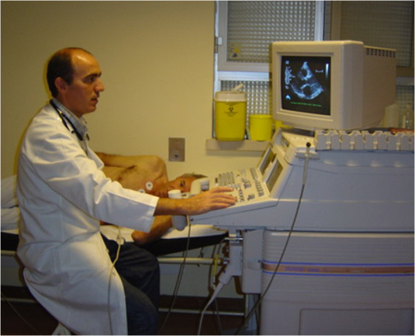
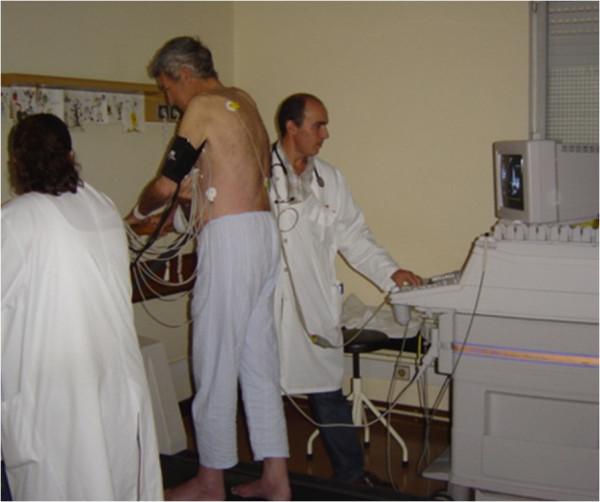
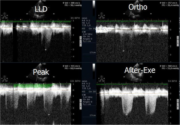
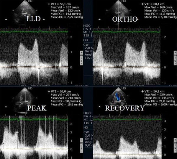
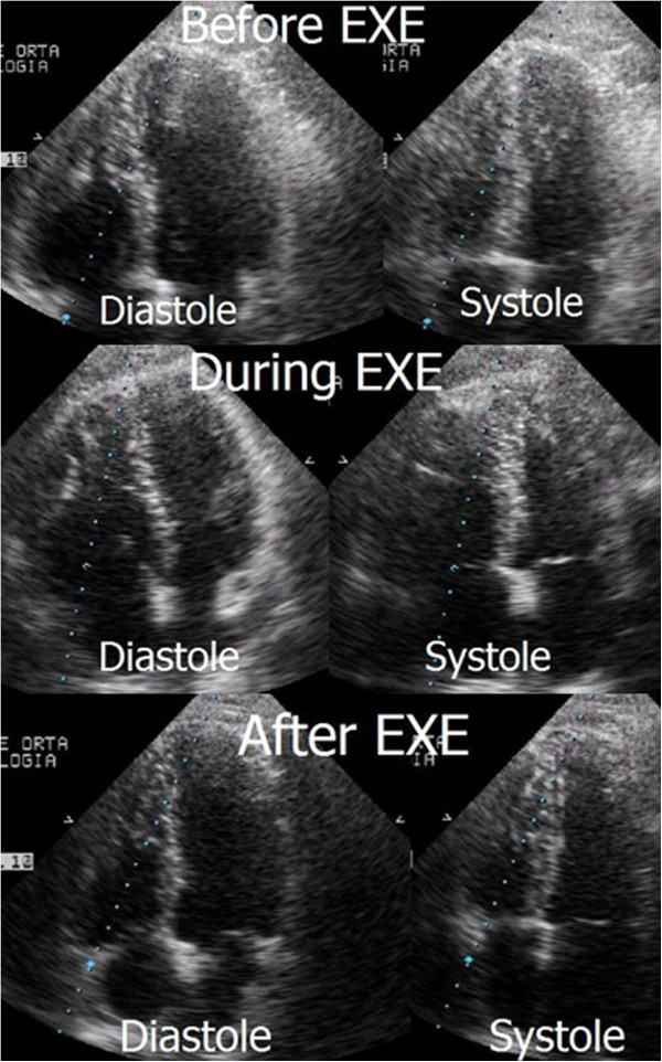
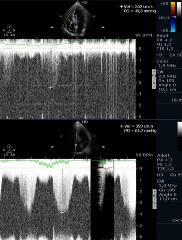
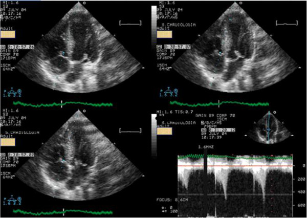
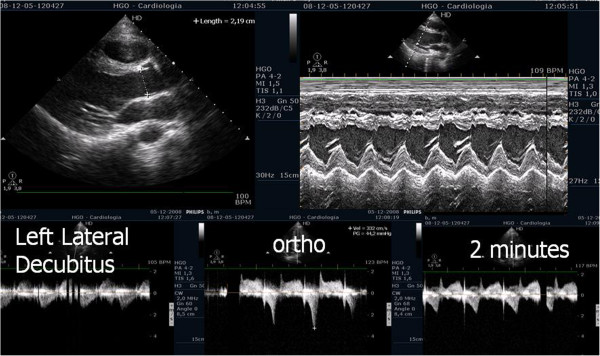
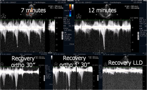
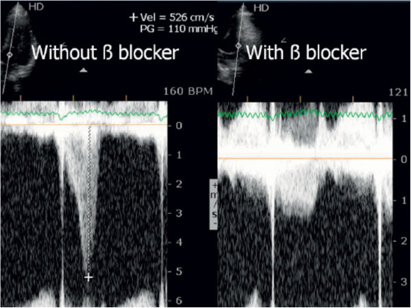
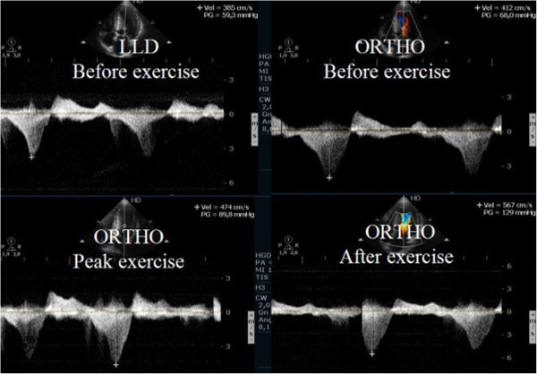
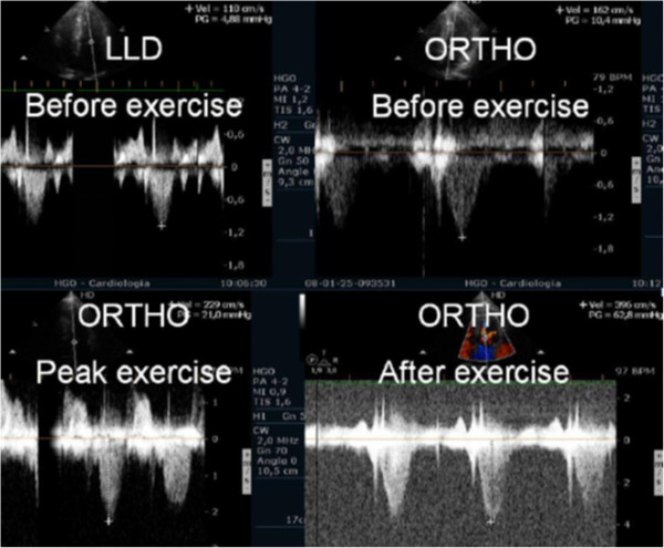
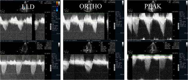
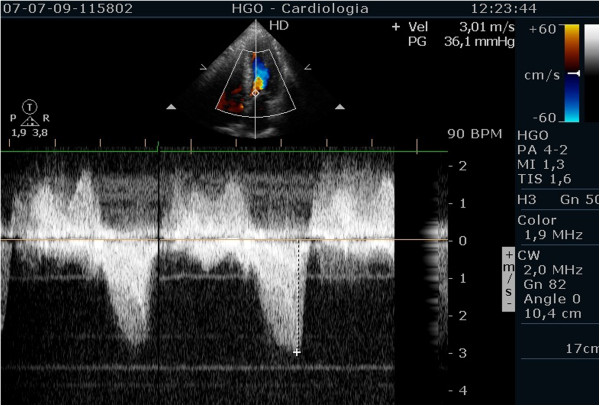
Exercise stress echocardiography is the most frequently used stress test in our laboratory. Exercise echocardiography is used mainly in the study of patients with coronary artery disease. However, the technique is increasingly being used to study other diseases. In our centre, we use an original methodology, published by us in 2000, in which we evaluate heart function during exercise in the treadmill. After the exercise, patients are maintained in orthostatic position when appropriate or lying down in left lateral decubitus for further evaluation. Since this method seems to increase the quality and the quantity of information obtained in so many clinical arenas, we now present a detailed review of this methodology and its applications.


Figures

Figure 1
Figure 1
Echocardiographic data acquisition with the patient in left lateral decubitus before exercise.
Figure 2
Figure 2
Echocardiographic data acquisition with the patient in orthostatic position during exercise in treadmill.
Figure 3
Figure 3
Right ventricular/ right atrium gradient, evaluated with CW Doppler, at different stages of the study in one patient with mitral stenosis.
Figure 4
Figure 4
Left atrium/ left ventricle gradient, evaluated with CW Doppler, at different stages of the study in one patient with mitral stenosis.
Figure 5
Figure 5
Apical four chamber view before, during, and after exercise with right ventricle dilatation only visible during exercise.
Figure 6
Figure 6
Continuous wave Doppler signal of the tricuspid regurgitant jet at peak exercise before and after injection of contrast.
Figure 7
Figure 7
Systolic anterior movement of mitral valve and significant intra-ventricular gradient detected at peak exercise.
Figure 8
Figure 8
Intra-ventricular gradient present in orthostatic position before exercise in one athlete decreases in the initial phase of exercise test.
Figure 9
Figure 9
Intra-ventricular gradient increases during the last part of exercise test and after exercise in orthostatic position. Obstruction suddenly disappears putting the athlete in decubitus.
Figure 10
Figure 10
IVG in one athlete assessed before and during beta blocker therapy.
Figure 11
Figure 11
Intra-ventricular gradient present in all the phases of the study in one patient increasing also after exercise in orthostatic position.
Figure 12
Figure 12
Intra-ventricular gradient present only in the recovery and in orthostatic position.
Figure 13
Figure 13
Intra-ventricular gradient present in all the phases of the study in one patient with symptomatic aortic stenosis increasing also after exercise in orthostatic position.
Figure 14
Figure 14
Intra-ventricular gradient evaluated in the same patient during beta blocker therapy.

References

    1. Wann LS, Faris JV, Childres RH, Dillon JC, Weyman AE, Feigenbaum H. Exercise cross-sectorial echocardiography in ischemic heart disease. Circulation. 1979;60:1300–1308. doi: 10.1161/01.CIR.60.6.1300. - DOI - PubMed
    1. Roger VL, Pellika PA, Oh JK, Miller FA, Sewward JB, Tajik AJ. Stress echocardiography. Part I. Exercise echocardiography: techniques, implementation, clinical applications, and correlations. Mayo Clin Proc. 1995;70:5–15. - PubMed
    1. Crouse LJ, Kramer PH. Exercise echocardiography: coming of age. J Am Coll Cardiol. 1994;24:115–116. doi: 10.1016/0735-1097(94)90550-9. - DOI - PubMed
    1. Armstrong WF. Exercise echocardiography: phase II, convincing the sceptics. J Am Coll Cardiol. 1992;19:82–83. doi: 10.1016/0735-1097(92)90055-R. - DOI - PubMed
    1. Medical Advisory Secretariat. Stress echocardiography for the diagnosis of coronary artery disease: an evidence-based analysis. Ont Health Technol Assess Ser. 2010;10(9):1–61. - PMC - PubMed