. 2014 Jan;128(1):140-51.
doi: 10.1111/jnc.12375. Epub 2013 Aug 26.

Synaptodendritic recovery following HIV Tat exposure: neurorestoration by phytoestrogens

Affiliations

Synaptodendritic recovery following HIV Tat exposure: neurorestoration by phytoestrogens

Sarah J Bertrand et al. J Neurochem. 2014 Jan.

Abstract

HIV-1 infects the brain and, despite antiretroviral therapy, many infected individuals suffer from HIV-1-associated neurocognitive disorders (HAND). HAND is associated with dendritic simplification and synaptic loss. Prevention of synaptodendritic damage may ameliorate or forestall neurocognitive decline in latent HIV-1 infections. The HIV-1 transactivating protein (Tat) is produced during viral latency in the brain and may cause synaptodendritic damage. This study examined the integrity of the dendritic network after exposure to HIV-1 Tat by labeling filamentous actin (F-actin)-rich structures (puncta) in primary neuronal cultures. After 24 h of treatment, HIV-1 Tat was associated with the dendritic arbor and produced a significant reduction of F-actin-labeled dendritic puncta as well as loss of dendrites. Pre-treatment with either of two plant-derived phytoestrogen compounds (daidzein and liquiritigenin), significantly reduced synaptodendritic damage following HIV-1 Tat treatment. In addition, 6 days after HIV-1 Tat treatment, treatment with either daidzein, or liquiritigenin enhanced recovery, via the estrogen receptor, from HIV-1 Tat-induced synaptodendritic damage. These results suggest that either liquiritigenin or daidzein may not only attenuate acute synaptodendritic injury in HIV-1 but may also promote recovery from synaptodendritic damage. The HIV-1 transactivating protein (Tat) is produced during viral latency in the brain. Treatment with either daidzein or liquiritigenin restored the loss of synaptic connectivity produced by HIV-1 Tat. This neurorestoration was mediated by estrogen receptors (ER). These results suggest that plant-derived phytoestrogens may promote recovery from HIV-1-induced synaptodendritic damage.

Keywords: F-actin; HAND; cell culture; daidzein; liquiritigenin; rat.


Conflict of interest statement

The authors have no conflicts of interest to declare.

Figures

Figure 1
Figure 1
Time-course of Tat-induced cell death and synaptodendritic alterations. A. The viability of primary cultures was assessed at 1, 24, 48, 72, 96 and 144 hours after treatment with Tat. There was no significant cell death through 24 hours; however, by 48 hours there was a significant decrease in cell viability (p<.001). By 72 hours, cell death reached a plateau and remained stable through 144 hours. Mean ± 95% confidence interval (CI). B. F-actin/MAP-2 staining of cell cultures at 1, 24, 72, and 144 hours after Tat 1-86B exposure. Tat 1-86B treatment produced a simplification of the fine network, as well as bundling and fragmentation of neurites at 24 hours, which preceded Tat-mediated cell death. Tat-induced alterations in the neuronal network persisted through 144 hours. C. Representative images (20×) of second branch order neuritic fragments co-stained with Oregon Green phalloidin and rabbit polyclonal anti-Tat primary/Alexa Red 594. Tat was associated with the F-actin labeled network at 1 hour and 24 hours post-treatment.
Figure 2
Figure 2
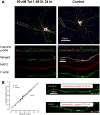
Computer-assisted identification of F-actin puncta after Tat treatment. A. Segments of second order dendritic branches (shown as boxes) were selected from 20× images of F-actin/MAP2/Hoechst-stained Tat-treated and non-treated control neurons. The computer-assisted intensity profile of F-actin fluorescence (green) showed numerous peaks corresponding to F-actin rich structures and valleys corresponding to low baseline staining of dendritic shafts. The computer-based intensity profile of MAP2-specific immunofluorescence (red) showed uniform labeling of all dendritic segments selected for counting of the F-actin puncta density. B. The total number of F-actin puncta (N; green +) were divided over the length of the neurite (L; green line) to provide the mean number of F-actin labeled puncta per 10μm of neuronal dendrite ± SEM. F-actin puncta were significantly reduced in neurons exposed to Tat for 24 hours using both computer-assisted intensity profiling and manual counting methods. F-actin puncta are readily distinguished and there was a very high correlation between the two methods (r2=.972).
Figure 3
Figure 3
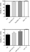
Pretreatment with phytoestrogens provides dose-dependent protection against neuronal cell death. A. Cell viability after DAI 24 hour pretreatment and a 48 hour incubation with 50nM Tat 1-86B. Treatment with 1μM DAI alone had no significant effect on cell viability relative to control cultures (F<1.0). DAI pretreatment provided complete neuroprotection in both the 0.2 μM and 1μM treatment groups (ps<0.001). Results are presented as mean % of control ± SEM. *Indicates significant protection of Tat-induced neurotoxicity. B. Cell viability after 24 hour LQ pretreatment and a 48 hour incubation with 50nM Tat 1-86B. There was no significant difference between LQ treated cultures and controls, indicating 1μM LQ was not neurotoxic. A linear dose-dependent effect of LQ was found (p<0.001) with significant, although not complete, neuroprotection. Results are presented as mean % of control ± SEM. *Indicates significant protection of Tat-induced neurotoxicity.
Figure 4
Figure 4
Pretreatment with DIA protects against Tat-induced loss of F-actin puncta. A. Chemical structure of daidzein (7,4′-Dihydroxyisoflavone). B. F-actin puncta following pretreatment (24 hours) with 1μM DAI and incubation (24 hours) with 50nM Tat 1-86 B. DAI was neither toxic nor stimulatory to production of F-actin puncta; however, Tat treated produced a significant loss of F-actin puncta (p<0.003), and DAI provided significant protection against the puncta loss caused by Tat. Results are presented as mean number of F-actin labeled puncta per 10 μm of neuronal dendrite ± SEM. *Indicates a significant loss of F-actin puncta after Tat 1-86B treatment when compared with vehicle treated controls (p<.01). **Indicates 1μM DAI pretreatment provided significant protection from damage induced by Tat 1-86B (50nM; 24 hours) when compared with cultures treated with Tat 1-86B alone. C-E. Neurons from (C) vehicle treated control cultures, (D) Tat 1-86B treated (50nM; 24 hours) cultures, and (E) pretreated DAI (1μM; 24 hours) + incubated with Tat 1-86B (50nM; 24 hours) cultures displaying typical F-actin (green), MAP-2 (red) and Hoescht (blue) staining for each treatment group. The control image shows robust F-actin presence, complex branching patterns, and an extensive fine network. Tat 1-86B treatment induced a simplification of the network. In contrast, in the DAI pre-treated culture, Tat 1-86B failed to cause network simplification, suggesting DAI pre-treatment protected against Tat induced synaptodendritic alterations.
Figure 5
Figure 5. Pretreatment with LQ provides protection against Tat-induced loss of F-actin puncta
A. Chemical structure of liquiritigenin (4′,7-Dihydroxyflavanone). B. Quantification of F-actin puncta with a pretreatment (24 hours) of 1μM LQ and 50nM Tat 1-86 B (24 hours). LQ did not alter F-actin puncta density from control (F<1.0), suggesting that LQ was neither toxic nor stimulatory. Tat treatment produced a significant loss of F-actin puncta (p<0.001) and LQ provided significant protection against the puncta loss caused by Tat (p<0.016). Results are presented as mean number of F-actin labeled puncta per 10 μm of neuronal dendrite ± SEM . *Indicates a significant loss of F-actin puncta after Tat 1-86B treatment when compared with vehicle treated controls (p<.01). **Indicates 1μM LQ pretreatment provided significant protection from F-actin puncta loss induced by Tat 1-86B (50nM; 24 hours) when compared with cultures treated with Tat 1-86B alone. C-E. Neurons from (C) vehicle treated control cultures, (D) Tat 1-86B treated (50nM; 24 hours) cultures, and (E) pretreated LQ (1μM; 24 hours) + incubated with Tat 1-86B (50nM; 24 hours) cultures, displaying typical F-actin (green), MAP-2 (red) and Hoescht (blue) staining for each treatment group. The control image shows robust F-actin presence, complex branching patterns, and an extensive fine network. Tat 1-86 treatment induced a simplification of the dendritic network. In contrast, in the LQ pre-treated culture, Tat 1-86B failed to cause network simplification, indicating LQ pre-treatment protected against Tat induced synaptodendritic alterations.
Figure 6
Figure 6
Phytoestrogens enhance the recovery of F-actin puncta from HIV-1 Tat-induced synaptodendritic injury in an estrogen-receptor dependent mechanism. The neurorecovery experiment was initiated by medium replacement after 6 days (i.e., after the initial cytotoxic effects of Tat). Three days after medium replacement (9 days after initial Tat treatment) a significant loss of F-actin puncta remained (p<0.001) in Tat-treated cultures. The cell cultures treated on day 6 with either DAI, LQ or TMX (tamoxifen) were not significantly different from controls. When either DAI or LQ were added on day 6 to the cultures initially treated with Tat, a significant increase in F-actin puncta was detected (p<0.002). This enhancement by DAI or LQ was blocked by the estrogen receptor antagonist TMX (100 nM), suggesting involvement of estrogen receptors in mediating the recovery of F-actin puncta. Results are presented as mean number of F-actin labeled puncta per 10 μm of neuronal dendrite ± SEM.

References

    1. Adams SM, Aksenova MV, Aksenov MY, Mactutus CF, Booze RM. ER-beta mediates 17beta-estradiol attenuation of HIV-1 Tat-induced apoptotic signaling. Synapse. 2010;64:829–838. - PMC - PubMed
    1. Adams SM, Aksenova MV, Aksenov MY, Mactutus CF, Booze RM. Soy Isoflavones Genistein and Daidzein Exert Anti-Apoptotic Actions via a Selective ER-mediated Mechanism in Neurons following HIV-1 Tat(1-86) Exposure. PLoS One. 2012;7:e37540. - PMC - PubMed
    1. Adle-Biassette H, Chretien F, Wingertsmann L, Hery C, Ereau T, Scaravilli F, Tardieu M, Gray F. Neuronal apoptosis does not correlate with dementia in HIV infection but is related to microglial activation and axonal damage. Neuropathol Appl Neurobiol. 1999;25:123–133. - PubMed
    1. Aksenov MY, Aksenova MV, Mactutus CF, Booze RM. Attenuated neurotoxicity of the transactivation-defective HIV-1 Tat protein in hippocampal cell cultures. Exp Neurol. 2009;219:586–590. - PMC - PubMed
    1. Aksenova MV, Aksenov MY, Adams SM, Mactutus CF, Booze RM. Neuronal survival and resistance to HIV-1 Tat toxicity in the primary culture of rat fetal neurons. Exp Neurol. 2009;215:253–263. - PMC - PubMed

Publication types

MeSH terms

Substances

LinkOut - more resources