Review
. 2014 May 20;20(15):2372-415.
doi: 10.1089/ars.2012.5147. Epub 2013 Oct 1.

SOD therapeutics: latest insights into their structure-activity relationships and impact on the cellular redox-based signaling pathways

Affiliations
Review

SOD therapeutics: latest insights into their structure-activity relationships and impact on the cellular redox-based signaling pathways

Ines Batinic-Haberle et al. Antioxid Redox Signal. .

Abstract

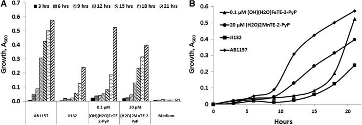
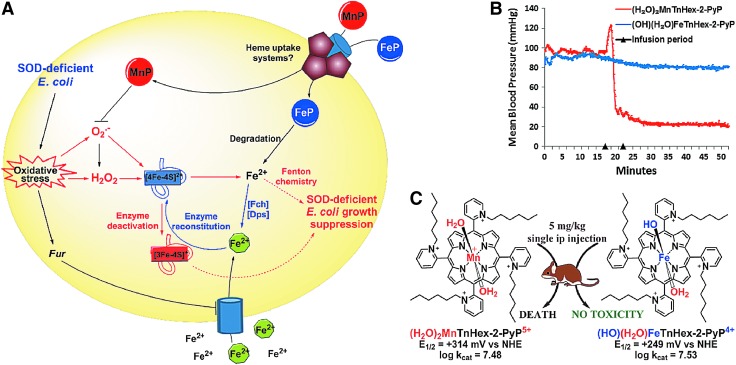
Significance: Superoxide dismutase (SOD) enzymes are indispensable and ubiquitous antioxidant defenses maintaining the steady-state levels of O2·(-); no wonder, thus, that their mimics are remarkably efficacious in essentially any animal model of oxidative stress injuries thus far explored.

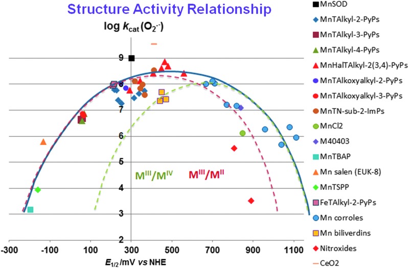
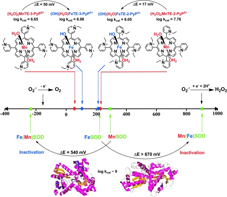
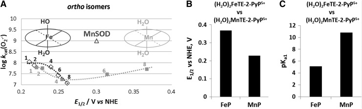
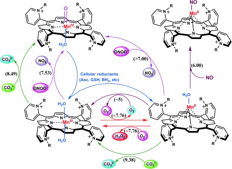
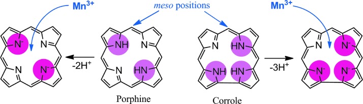
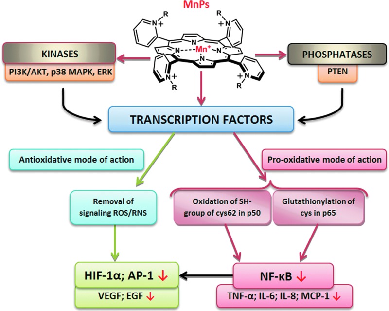
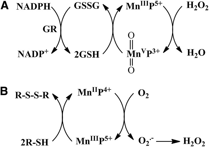
Recent advances: Structure-activity relationship (half-wave reduction potential [E1/2] versus log kcat), originally reported for Mn porphyrins (MnPs), is valid for any other class of SOD mimics, as it is dominated by the superoxide reduction and oxidation potential. The biocompatible E1/2 of ∼+300 mV versus normal hydrogen electrode (NHE) allows powerful SOD mimics as mild oxidants and antioxidants (alike O2·(-)) to readily traffic electrons among reactive species and signaling proteins, serving as fine mediators of redox-based signaling pathways. Based on similar thermodynamics, both SOD enzymes and their mimics undergo similar reactions, however, due to vastly different sterics, with different rate constants.

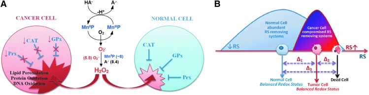
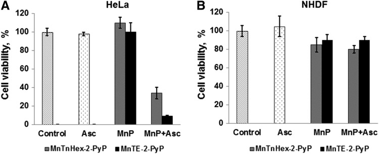
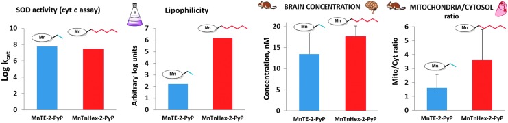
Critical issues: Although log kcat(O2·(-)) is a good measure of therapeutic potential of SOD mimics, discussions of their in vivo mechanisms of actions remain mostly of speculative character. Most recently, the therapeutic and mechanistic relevance of oxidation of ascorbate and glutathionylation and oxidation of protein thiols by MnP-based SOD mimics and subsequent inactivation of nuclear factor κB has been substantiated in rescuing normal and killing cancer cells. Interaction of MnPs with thiols seems to be, at least in part, involved in up-regulation of endogenous antioxidative defenses, leading to the healing of diseased cells.

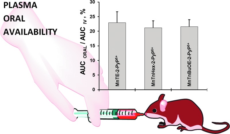
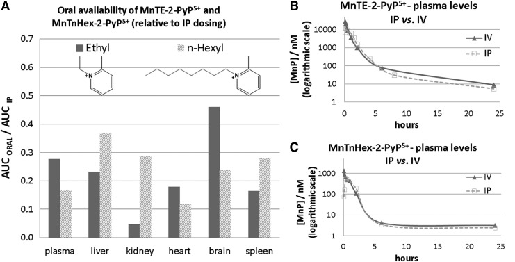
Future directions: Mechanistic explorations of single and combined therapeutic strategies, along with studies of bioavailability and translational aspects, will comprise future work in optimizing redox-active drugs.


Figures

<b>FIG. 1.</b>
FIG. 1.
Main classes of “true” SOD mimics: compounds that catalyze O2· dismutation (oxidize and reduce O2·). The catalysis of dismutation should occur with kcat higher than k for O2· self-dismutation of ∼5×10⁵ M−1s−1 at pH 7 (109). Shown are the structures of optimized MnP, Mn corrole, Mn cyclic polyamine, and Mn salen. Cerium dioxide comes in a form of ceria nanoparticles. OsO4 is too toxic for therapeutic purposes regardless of its kcat as high as that of SOD enzyme. Mn2+ is a fair SOD mimic. It has not been used much in preclinical research perhaps due to the toxicity described as manganism (15, 118, 230). MnP, Mn porphyrin; SOD, superoxide dismutase.
<b>FIG. 2.</b>
FIG. 2.
The design of MnP-based redox-active drugs. Accomplished in three phases, it resulted in the creation of three lead compounds: MnTE-2-PyP5+, MnTnHex-2-PyP5+, and MnTnBuOE-2-PyP5+ (216, 271). In the first phase, the major benefit of the ortho- positioned quaternary nitrogens imposing a strong electro-withdrawing effect on Mn site was demonstrated. In phase 2, the lipophilicity was increased a few orders of magnitude by lengthening the N-alkylpyridyl chains from ethyl to n-octyl; the longer the chains are, the higher the compound bioavailability and, in turn, therapeutic efficacy of the compound. In phase 3, oxygens were introduced deep into the alkyl chains. Such a compound, MnTnBuOE-2-PyP5+ is approximately four to five-fold less toxic to mouse than MnTnHex-2-PyP5+ (26, 216, 271).
<b>FIG. 3.</b>
FIG. 3.
The toxicity of different Mn(III) N-substituted pyridylporphyrins. (A) Comparison of Mn(III) N-butoxyethylpyridylporphyrin to its n-hexyl and n-heptyl analogs when given via a single ip injection. (B) Comparison of their efficacy and toxicity in aerobic growth of SOD-deficient (sod1Δ) yeast S. cerevisiae (EG118) relative to wild-type yeast (EG 103). Cultures in 96-well plates were grown aerobically at 30°C and 250 rpm on a thermostatic shaker in peptone agar supplemented with 2% dextrose (216). ip, intraperitoneal.
<b>FIG. 4.</b>
FIG. 4.
Structure-activity relationships (SARs) correlate the redox potency expressed as one-electron E1/2, and the ability of compounds to catalyze the O2· dismutation, log kcat(O2·). With Mn(III) biliverdins and Mn(III) corroles, MnIV/MnIII redox couple is involved in the catalysis of O2· dismutation; with all other Mn compounds, the MnIII/MnII redox couple is involved. The E1/2 for nitroxides relates to the oxidation of nitroxide to oxoammonium cation (14, 167, 228). The SAR (showed with thick blue line) is valid for many redox-active compounds regardless of their structure: metalloporphyrins, Mn(IV) biliverdins, Mn(III) salens, Mn(II) polyamines, Mn(III) corroles, and Mn(II) aqua complex (271). The E1/2 values for MnIVC/MnIIIC redox couple (given in Table 1) were determined in phosphate buffer for anionic Mn corrole with two sulfonatopyrrolic groups and three meso pentaphenyl groups, but in acetonitrile for cationic Mn corrole, which bears two meso para-methylpyridyl groups and one meso substituent with derivatized tetrapfluorophenyl group (80, 104, 196) (Fig. 1). Thus, it is not straighforward to predict the magnitude of the shift in E1/2 from one to another corrole. According to Marcus equation, the 120 mV shift in E1/2 should cause a 10-fold change in rate constant (29). The increase in log kcat(O2·) for ∼3 orders of magnitude (from Mn corrole) should have shifted the E1/2 for ∼360 mV, from ∼+840 mV vs. Ag/AgCl (+1040 mV vs. NHE) to ∼+500 mV versus Ag/AgCl (+700 mV vs. NHE) (Fig. 5). When translated into experimental data, it appears that E1/2 for these compounds is identical in either solvent: water or acetonitrile (80, 104, 196). Below the SAR, indicated as a thick blue line, the two individual SARs relate to complexes that catalyze O2· dismutation employing either MnIII/MnII (red line) or MnIV/MnIII redox couple (green line). The optimal reduction potential (peak) of these SARs differs ∼300 mV. The compounds with very negative and very positive values of E1/2 are essentially unable to dismute O2·; different strategies have been employed to improve their E1/2. The electron-withdrawing groups are needed for the majority of metallporphyrins to move their potential from negative values into the region of optimal E1/2 values (+200 to +400 mV vs. NHE) (28). However, with Mn corroles, the electron-donating groups are required for modifying their E1/2 from ∼+1000 to ∼+700 mV versus NHE (196). Since their redox is irreversible, the nitroxides do not fit the SAR well (red rhombi). Any major deviation from SAR indicates that factors other than thermodynamics have an impact on the kcat (27, 28, 221, 223). Charges are omitted in the figure legend for simplicity. E1/2, half-wave reduction potential; NHE, normal hydrogen electrode.
<b>FIG. 5.</b>
FIG. 5.
Comparison of the effect of the type of metal on the SOD activity of enzymes and metalloporphyrins. Replacing the metal sites between MnSOD and FeSOD enzymes precludes the appropriate amino-acid configuration of metal site and, thus, results in vastly shifted E1/2 out of the limits required for superoxide dismutation; consequently, the enzyme becomes inactive. However, when different metals are ligated to the same porphyrin, the change in the type of axial ligand is easily achievable: Fe hydroxo axial versus Mn axial water at pH 7.8 (sixth coordination site in these complexes is occupied by a water molecule). In such structures, both Mn aqua and Fe hydroxo porphyrins have essentially identical E1/2 and, therefore, similar SOD-like activity. To see this illustration in color, the reader is referred to the web version of this article at www.liebertpub.com/ars
<b>FIG. 6.</b>
FIG. 6.
SAR (A) and thermodynamic parameters (B, C) for the series of ortho Fe(III) N-alkylpyridylporphyrins as compared with Mn analogs (272, 273). The different shape of SAR is due to the difference between the chemistry of Fe and MnPs. At pH 7.8, FePs bear one hydroxo ligand that neutralizes single charge on Fe and labilizes trans-axial water, which, in turn, impacts the kcat and E1/2. To stress the difference, we showed here also the electrochemical data on both Fe and MnPs bearing same axial ligands—water molecules—and therefore same charge, but their E1/2 differ ∼150 mV; we have shown earlier that the effect, which impacts the E1/2, also affects the strength of the M-H2O bond. The more electron-deficient the metal complex, the higher the E1/2 it is, and the stronger it binds the axial water; consequently, the loss of its proton is promoted, which is reflected in lower pKa1 (84, 86, 221). FePs, Fe porphyrins.
<b>FIG. 7.</b>
FIG. 7.
Comparison of ortho Fe(III) and Mn(III) N-ethylpyridylporphyrins in protecting SOD-deficient Escherichia coli while growing aerobically. FeP allows E. coli to overgrow the wild type at ∼1000-fold lower concentration than MnP (A). However, the growth pattern is different (B). The growth was followed in a restricted five-amino-acid medium that better distinguishes the true SOD mimics from other redox-active compounds than rich media (270, 273). Wild-type E. coli was AB1157, and SOD-deficient JI132.
<b>FIG. 8.</b>
FIG. 8.
FePs and MnPs differ greatly with respect to their chemistry which translates to differences in their biology. The schematic presentation of differences in the in vivo protective (A) and toxic (B, C) effects of FePs and MnPs on SOD-deficient E. coli. The (H2O)2MnTE-2-PyP5+ and (H2O)(OH)FeTE-2-PyP5+ have very similar E1/2 (Table 1) and similar electrostatics and, in turn, fairly similar kcat(O2·). Consequently, FePs and MnPs should protect SOD-deficient E. coli when growing aerobically to a similar extent and at similar concentrations—some difference may be due to the difference in the total charge of these two classes of compounds. However, 20 μM MnPs was fully protective; while 20 μM FePs was toxic. On uptake, FePs undergo rapid degradation with H2O2 produced during fast redox cycling with cellular reductants (ascorbate or thiols), whereby “free” Fe2+ is released. The iron-transporting/sequestering siderophores (Fch—ferrochelatase; Dps—Fe-storage protein; green circles) take care of “free” Fe. At very low levels (0.01 to 1 μM), Fe2+ could reconstitute Fe-containing enzymes, such as aconitases, threonine dehydrogenase, ribulose-5-phosphate 3-epimerase, and peptide deformylase (69). These enzymes undergo superoxide-driven oxidative degradation and subsequent reversible release of Fe2+. At higher concentrations, the deleterious effects of iron-mediated Fenton chemistry-driven·OH radical production may prevail. The MnPs are much more resistant toward oxidative degradation and, thus, as SOD mimics, they eliminate superoxide, thereby preventing superoxide-driven damage on Fe-containing enzymes. A single ip injection of (H2O)2MnTnHex-2-PyP5+ caused mouse death, while no toxicity was seen with Fe analogue. For reasons not presently understood, H2O)2MnTnHex-2-PyP5+, but not (OH)(H2O)FeTnHex-2-PyP4+ causes blood pressure drop. Modified from Tovmasyan et al. (273). To see this illustration in color, the reader is referred to the web version of this article at www.liebertpub.com/ars
<b>FIG. 9.</b>
FIG. 9.
The reactivities of ortho isomers of Mn(III) N-substituted pyridylporphyrins toward small molecules. R describes either alkyl or alkoxyalkyl groups. The reactivity toward protein thiols has not been included in this scheme, but is discussed under Reactivity toward cellular reductants and Reactivity toward signaling proteins sections. The reactivity is predominantly determined by the presence of cationic charges on nitrogens that dominate the electronics and electrostatics of the reactions. The removal of O2· or ONOO, resulting in H2O2 or O=MnIVP production, can only bear antioxidative character if the cell has either sufficient peroxide-removing enzymes or reductants to eliminate strong oxidants (high valent metal), respectively. If not, as is often the case with oxidative stress injuries and in a particular cancer, the pro-oxidative action may prevail. Under such conditions, MnP may employ H2O2 to inhibit the activation of NF-κB by oxidizing and/or glutathionylating its subunits (124, 125). Moreover, MnP may directly oxidize thiols (see below under Thiols section). NF-κB, nuclear factor κB. To see this illustration in color, the reader is referred to the web version of this article at www.liebertpub.com/ars
<b>FIG. 10.</b>
FIG. 10.
The comparison of porphyrin versus corrole core. Porphyrin contains 2 while corrole contains 3 protonated pyrrolic nitrogens (encircled). Consequently, upon deprotonation porphyrin is a dianionic, while corrole is a trianionic ligand. Such a difference results in differential metal/ligand stability, and affects properties of metals. To see this illustration in color, the reader is referred to the web version of this article at www.liebertpub.com/ars
<b>FIG. 11.</b>
FIG. 11.
Differential effect of MnP/ascorbate-driven H2O2 production on tumor and normal cells. (A) Ascorbate oxidation produces H2O2 that normal cells remove readily. Depending on the type of cancer cell, peroxide may not be well taken care of, leading to higher oxidative stress than in normal cells (16) (B) Redox status of cancer and tumor cell differs significantly, which determines their differential sensibility to an additional increase in reactive species. Tumor cells already have high levels of reactive species, and any further increase could cause their death (42, 114). To see this illustration in color, the reader is referred to the web version of this article at www.liebertpub.com/ars
<b>FIG. 12.</b>
FIG. 12.
Differential cytotoxicity of MnP/ascorbate in tumor (A) and normal cell line (B). Two MnPs were tested, a hydrophilic MnTE-2-PyP5+ and a lipophilic MnTnHex-2-PyP5+ at 3 μM (shown) and 15 μM. The concentration of ascorbate was 1 mM. MnPs and ascorbate alone were not toxic. In the presence of ascorbate, both MnPs become toxic to cervical cancer cells, HeLa, but not to normal primary fibroblasts, NHDF cells. MTT assays were performed in a 96-well plate with an initial seeding density of 25,000 cells/well. Cells were incubated with MnP ±ascorbate for 48 h before the assay was performed (272, 299). The study was done in duplicate. Ascorbate was added soon after MnP. The effect of MnP/ascorbate on several other cancer cell lines has also been demonstrated (82, 299).
<b>FIG. 13.</b>
FIG. 13.
The impact of MnPs on transcription factors and kinases and phosphatases and, in turn, on the related genes. To see this illustration in color, the reader is referred to the web version of this article at www.liebertpub.com/ars
<b>FIG. 14.</b>
FIG. 14.
The effect of MnTnHex-2-PyP5+(A) on suppression of stroke injury in a mouse middle cerebral artery occlusion stroke model (238). (B) Infarct volumes were measured 7 days after 90 min MCAO. Rats were treated with intravenous vehicle (0.3 ml phosphate-buffered saline) or MnTnHex-2-PyP5+ (225 μg/kg) 5 min after reperfusion onset. The doses were repeated twice daily as subcutaneous injections for 7 days, after which cerebral infarct volume was measured. Open circles indicate individual animal values. Horizontal lines indicate group median values. MnTnHex-2-PyP5+ reduced cerebral infarct volume in the cortex (p=0.05) and subcortex (p=0.01), which was reflected in a 32% reduction in total infarct volume (p=0.028). Open circles indicate individual animal values. Horizontal lines indicate group mean values. (C) In studies on cytokines, rats were subjected to 90 min of middle cerebral artery occlusion. Five minutes after onset of reperfusion, rats were randomly treated with vehicle (n=3) or 225 μg/kg IV MnTnHex-2-PyP (n=3) followed by subcutaneous vehicle or 225 μg/kg MnTnHex-2-PyP5+, respectively, at 12 and 18 h post-MCAO. Brains were harvested at 24 h post-MCAO and analyzed for TNF-α and IL-6 by fluorescent enzyme-linked immunosorbent assay. Values represent mean±s.d. Both TNF-α and IL-6 concentrations were decreased by MnTnHex-2-PyP5+ (*p<0.05). (D) In NF-κB studies, four rats were subjected to 90 min middle cerebral artery occlusion and then treated with vehicle or MnTnHex-2-PyP5+ (225 μg/kg IV). Six hours later, ischemic brain was harvested to obtain nuclear extracts (2.5 μg) for electromobility shift assay (EMSA). The intravenous MnTnHex-2-PyP5+ decreased postischemic NF-κB DNA binding to a κB consensus oligo. Data shown are from Upper gel (EMSA): A-C are control lanes (A=probe only, B=positive control [HeLa nuclear extract], C=cold competitor). D and E (without and with p65 antibody, respectively) relate to rat #1 (vehicle). F and G (without and with p65 antibody, respectively) relate to rat #2 (MnTnHex-2-PyP5+). H and I=vehicle rat #3 (with and without p65). J and K=rat #4 (MnTnHex-2-PyP5+) with and without p65. Two slower migrating DNA-binding complexes are observed (shift). The proteins in the slower migrating complexes were identified by supershift analysis with 1 μg of p65-specific antibody. A marked reduction in NF-κB binding is seen in rats #2 and #4 (lanes F, G, J, and K, both hexyl). MCAO, middle cerebral artery occlusion. To see this illustration in color, the reader is referred to the web version of this article at www.liebertpub.com/ars
<b>FIG. 15.</b>
FIG. 15.
The anticancer action of MnP as a sole agent. Anticancer effects of MnTE-2-PyP5+ (A) in a mouse sc 4T1 xenograft breast cancer model. 4T1 murine breast tumors were grown in Balb/C mice, allowed to reach ≥200 mm3 in size, and randomized to one of the three treatment groups: PBS; 2×1 mg/kg/day MnTE-2-PyP5+ (low) and 2×7.5 mg/kg/day MnTE-2-PyP5+ (high). MnP was given sc throughout the duration of the study. Immunohistochemistry of HIF-1α (C), carbonic anhydrase CAIX (D), and pimonidazole (E) and representative Western blot for HIF-1α is shown (B). Densitometric readings (HIF-1α/α-tubulin) of the Western blot are expressed as percentage control. While significant changes in different signaling proteins that impact the tumor growth were demonstrated, the effect on tumor growth delay was significant but not large, even at such a high dose as 15 mg/kg/day (213). HIF-1α, hypoxia inducible factor-1α. To see this illustration in color, the reader is referred to the web version of this article at www.liebertpub.com/ars
<b>FIG. 16.</b>
FIG. 16.
The chemosensitizing effect of MnE-2-PyP5+ in cellular lymphoma study (125). Dexamethasone alone (1 μM) and MnTE-2-PyP5+ (50 nM) each increased levels of reactive species, which resulted in glutathionylation of p65 and subsequent suppression of NF-κB DNA binding. The effects are largely enhanced with their co-administration to murine thymic lymphoma WEHI7.2 cells maintained in suspension in DMEM+10% calf serum. DMEM, Dulbecco's Modified Eagle Medium-low glucose. To see this illustration in color, the reader is referred to the web version of this article at www.liebertpub.com/ars
<b>FIG. 17.</b>
FIG. 17.
The glutathione peroxidase (GPx) and thiol oxidase (TO) activities may contribute to the biological actions of MnPs. The proposed GPx-like (A) and thiol oxidase (B) activities of MnPs. The scheme is proposed based on the aqueous chemistry and experimental data in cellular and animal studies (272).
<b>FIG. 18.</b>
FIG. 18.
Comparison of the SOD-like potency, logkcat(O2·), lipophilicity, and accumulation in mitochondria and brain for two MnPs. MnTE-2-PyP5+ and MnTnHex-2-PyP5+ have similar redox-based properties, identical charge, and are, thus, among the most potent SOD mimics and peroxynitrite reductants. However, they differ greatly with regard to lipophilicity, bulkiness, and shape. These differences translate into differences in their bioavailability and, in turn, therapeutic potential. In mouse studies, MnPs were given to C57BL6 mice sc for 5 days, twice daily at 2 mg/kg. MnP levels were measured in brain (289) and in heart mitochondria and cytosol at 6 h after the last injection (250, 254). To see this illustration in color, the reader is referred to the web version of this article at www.liebertpub.com/ars
<b>FIG. 19.</b>
FIG. 19.
Plasma oral availability of three water-soluble but differently hydrophilic Mn(III) N-substituted pyridylporphyrins, MnTE-2-PyP5+, MnTnHex-2-PyP5+, and MnTnBuOE-2-PyP5+. The unpublished data (Spasojevic et al.) for MnTnBuOE-2-PyP5+ are also shown. The data for MnTE-2-PyP5+ and MnTnHex-2-PyP5+ are from Ref. (288). To see this illustration in color, the reader is referred to the web version of this article at www.liebertpub.com/ars
<b>FIG. 20.</b>
FIG. 20.
Organ oral availability of two water-soluble, differently hydrophilic Mn(III) N-alkylpyridylporphyrins, MnTE-2-PyP5+ and MnTnHex-2-PyP5+. (A) The AUCIP was taken as 100% (1 unit). The plasma, liver, kidney, heart, brain, and spleen AUCORAL/AUCIP values for MnTE-2-PyP5+ and MnTnHex-2-PyP5+. MnTE-2-PyP5+ was given orally at 10 mg/kg, while MnTnHex-2-PyP5+ at 2 mg/kg. The plasma AUCIP was found to be (B) 83% of AUCIV for MnTE-2-PyP5+ and (C) 84% for MnTnHex-2-PyP5+. At a 5-fold dose difference, the average distribution of drug in organs is either similar or higher with hexyl than with ethyl species (288). Only pyridyl substituents at meso positions of porphyrin ring are shown.
<b>FIG. 21.</b>
FIG. 21.
Differential accumulation of MnPs in tumor and normal tissue. (A) T2-weighted image of prostate cancer with hind flank muscle (Top) of C57Bl mice. Overlay of T1-weighted image over T2-weighted image (Bottom-Left, predose and Bottom-Right, post ip) shows contrast enhancement within the tumor (in vivo RM-9 model) after 8 mg/kg ip administration of MnTE-2-PyP5+ (183). (B) The accumulation of MnTnHex-2-PyP5+ measured by LCMS/MS in 4T1 breast cancer sc xenograft tumor and in the muscle from the opposite leg when MnP was given as a single agent (twice daily sc at 1 mg/kg), or in the presence of ascorbate (twice daily ip at 2 g/kg for the duration of study) (299). Ascorbate did not affect MnP accumulation. LCMS, liquid chromatography-tandem mass spectrometry.
<b>FIG. 22.</b>
FIG. 22.
The effect of MnTDE-2-ImP5+ on oxidative stress and, in turn, on PTEN signaling pathways involved in a mouse pulmonary radioprotection. Shown are changes in PTEN signaling as measured by PTEN expression and protein levels obtained by immunostaining; the same finding was supported with Western blotting. Also shown are protein levels of Bax and p53 (western blotting). PI3K-AKT is a key signaling pathway that is negatively regulated by PTEN. Finally, the changes in transforming growth factor expression (TGF)-β1 (by immunostaining), NOX4 expression and protein levels (by immunostaining, and Western blotting), and 8-OHdG levels (by immunostaining) are also presented (302). 8-OHdG, 8-oxo-2′-deoxyguanosine; PTEN, phosphoinositide 3-phosphatase. To see this illustration in color, the reader is referred to the web version of this article at www.liebertpub.com/ars
<b>FIG. 23.</b>
FIG. 23.
MnP protects against prostate radiation-induced injury to male reproductive system. The reversal of erectile dysfunction and the reduction in testes shrinkage by MnTE-2-PyP5+, induced after rat prostate radiation (A–C) as a consequence of radiation-induced oxidative stress (D–F) (192). Intracavernous pressure (ICP) was obtained after cavernous nerve stimulation as a measurement of erectile function 12 weeks postirradiation. Irradiation caused a significant decrease in ICP (RAD group) (B) as compared with the nonirradiated group (PBS). MnTE-2-PyP5+ protected from the irradiation-induced loss in ICP (MnTE-2-PyP RAD). n=8 rats/group, “*” denotes a significant difference from PBS group, p=0.05, and “#” denotes a significant difference from RAD group, p=0.05. The MnP also prevented testes shrinkage (C), hair loss (not shown), and the damage to prostate tissue (not shown) (192). The oxidative stress is demonstrated as the increase in NAPDH oxidase expression (F), macrophage infiltration (ED-1) (E), and Nrf-2 (the primary cellular defense against the cytotoxic effects of oxidative stress) up-regulation (D) (136) Nrf-2, nuclear factor-erythroid-derived 2-like 2. To see this illustration in color, the reader is referred to the web version of this article at www.liebertpub.com/ars
<b>FIG. 24.</b>
FIG. 24.
The radiosensitizing effect of a lipophilic MnTnHex-2-PyP5+ in a sc xenograft nu/nu mouse Balb/c D 245-MG glioblastoma multiform model (21, 22). The mice (8/group) were treated twice daily via sc injections of 1.6 mg/kg of MnP (starting at 24 h before radiation and continued during the duration of the study), or 1 Gy radiation (3 days 1 Gy per day) and their combination. To see this illustration in color, the reader is referred to the web version of this article at www.liebertpub.com/ars

References

    1. Abashkin YG. and Burt SK. (salen)MnIII compounds as nonpeptidyl mimics of catalase. Mechanism-based tuning of catalase activity: a theoretical study. Inorg Chem 44: 1425–1432, 2005 - PubMed
    1. Abraham SK, Sarma L, and Kesavan PC. Protective effects of chlorogenic acid, curcumin and beta-carotene against gamma-radiation-induced in vivo chromosomal damage. Mutat Res 303: 109–112, 1993 - PubMed
    1. Aitken JB, Shearer EL, Giles NM, Lai B, Vogt S, Reboucas JS, Batinic-Haberle I, Lay PA, and Giles GI. Intracellular targeting and pharmacological activity of the superoxide dismutase mimics MnTE-2-PyP5+ and MnTnHex-2-PyP5+ regulated by their porphyrin ring substituents. Inorg Chem 52: 4121–4123, 2013 - PubMed
    1. Allen BG, Sibenaller ZA, Cullen JJ, Buettner GR, Welsh JJ, Wagner BA, van't Erve TJ, Buatti JM, Carlisle TL, Smith MC, Walsh SA, Bayouth JE, TenNapel M, and Spitz DR. Pharmacological ascorbate enhances chemo-radio-sensitization in brain and lung cancer. Free Radic Biol Med 53: S39, 2012
    1. Andrievsky GV, Bruskov VI, Tykhomyrov AA, and Gudkov SV. Peculiarities of the antioxidant and radioprotective effects of hydrated C60 fullerene nanostuctures in vitro and in vivo. Free Radic Biol Med 47: 786–793, 2009 - PubMed

Publication types

MeSH terms