

Genes related to suppression of malignant phenotype induced by Maitake D-Fraction in breast cancer cells
- PMID: 23875900
- PMCID: PMC3719462
- DOI: 10.1089/jmf.2012.0222
Genes related to suppression of malignant phenotype induced by Maitake D-Fraction in breast cancer cells
Abstract
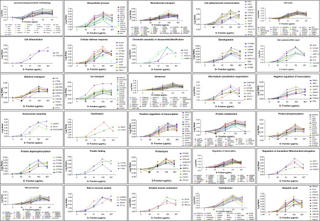
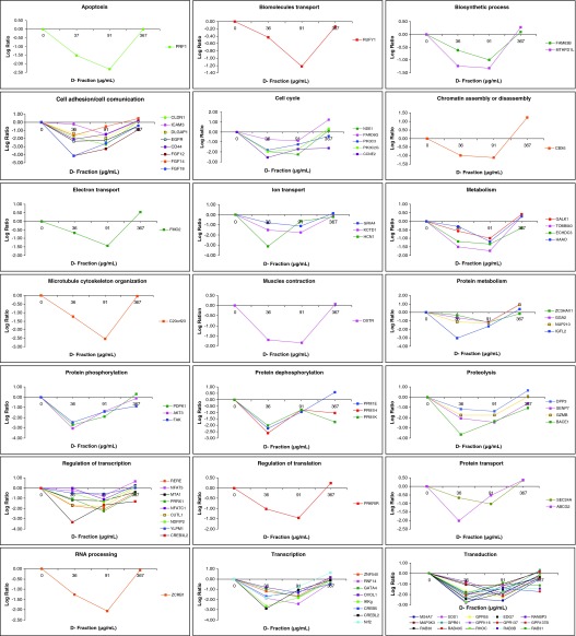
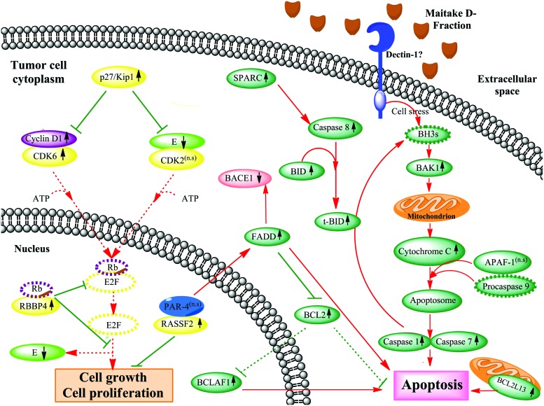
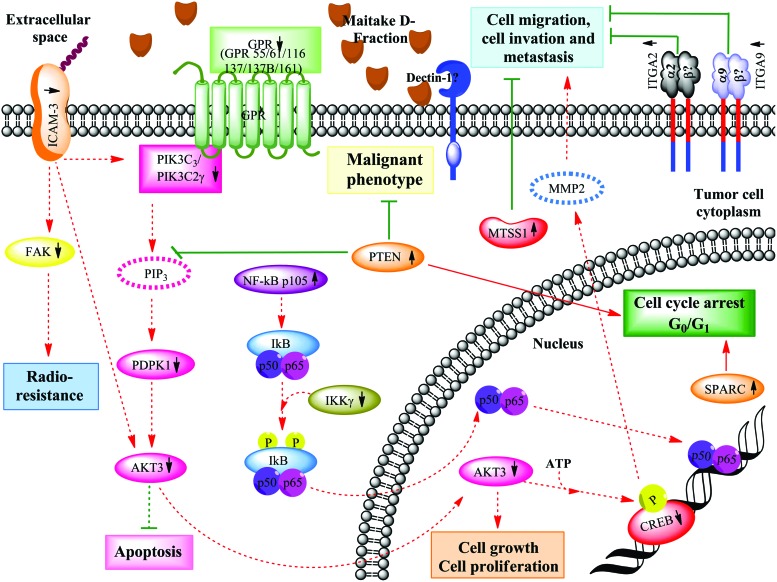
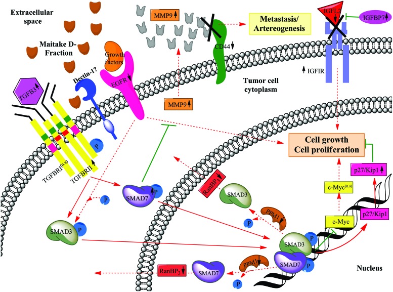
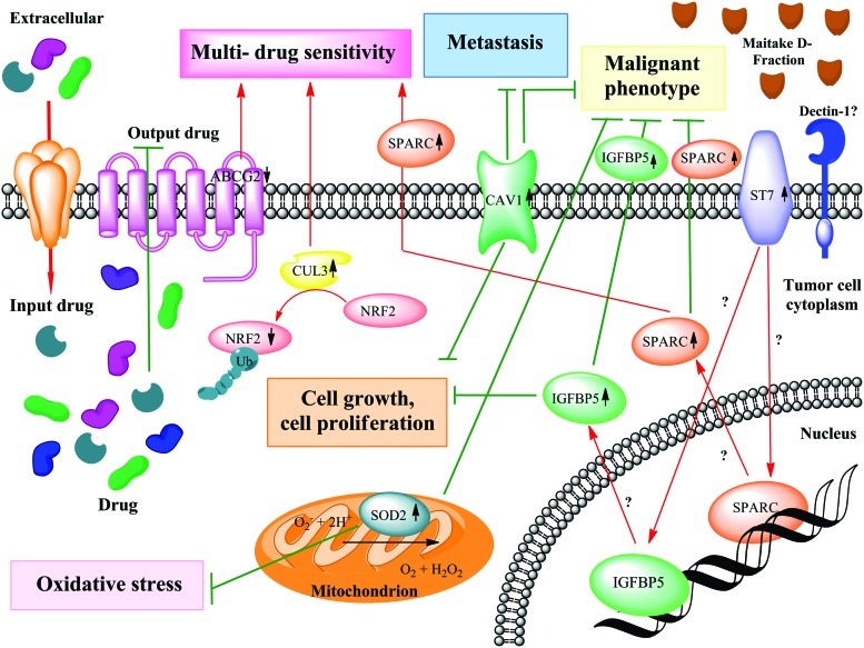
It is already known that the Maitake (D-Fraction) mushroom is involved in stimulating the immune system and activating certain cells that attack cancer, including macrophages, T-cells, and natural killer cells. According to the U.S. National Cancer Institute, polysaccharide complexes present in Maitake mushrooms appear to have significant anticancer activity. However, the exact molecular mechanism of the Maitake antitumoral effect is still unclear. Previously, we have reported that Maitake (D-Fraction) induces apoptosis in breast cancer cells by activation of BCL2-antagonist/killer 1 (BAK1) gene expression. At the present work, we are identifying which genes are responsible for the suppression of the tumoral phenotype mechanism induced by Maitake (D-Fraction) in breast cancer cells. Human breast cancer MCF-7 cells were treated with and without increased concentrations of Maitake D-Fraction (36, 91, 183, 367 μg/mL) for 24 h. Total RNA were isolated and cDNA microarrays were hybridized containing 25,000 human genes. Employing the cDNA microarray analysis, we found that Maitake D-Fraction modified the expression of 4068 genes (2420 were upmodulated and 1648 were downmodulated) in MCF-7 breast cancer cells in a dose-dependent manner during 24 h of treatment. The present data shows that Maitake D-Fraction suppresses the breast tumoral phenotype through a putative molecular mechanism modifying the expression of certain genes (such as IGFBP-7, ITGA2, ICAM3, SOD2, CAV-1, Cul-3, NRF2, Cycline E, ST7, and SPARC) that are involved in apoptosis stimulation, inhibition of cell growth and proliferation, cell cycle arrest, blocking migration and metastasis of tumoral cells, and inducing multidrug sensitivity. Altogether, these results suggest that Maitake D-Fraction could be a potential new target for breast cancer chemoprevention and treatment.
Figures
Similar articles
-
Maitake (D fraction) mushroom extract induces apoptosis in breast cancer cells by BAK-1 gene activation.J Med Food. 2011 Jun;14(6):563-72. doi: 10.1089/jmf.2010.0095. Epub 2011 Apr 11. J Med Food. 2011. PMID: 21480800
-
Maitake Pro4X has anti-cancer activity and prevents oncogenesis in BALBc mice.Cancer Med. 2016 Sep;5(9):2427-41. doi: 10.1002/cam4.744. Epub 2016 Jul 11. Cancer Med. 2016. PMID: 27401257 Free PMC article.
-
Antitumoral Effects of D-Fraction from Grifola Frondosa (Maitake) Mushroom in Breast Cancer.Nutr Cancer. 2017 Jan;69(1):29-43. doi: 10.1080/01635581.2017.1247891. Epub 2016 Nov 28. Nutr Cancer. 2017. PMID: 27892708
-
Can maitake MD-fraction aid cancer patients?Altern Med Rev. 2002 Jun;7(3):236-9. Altern Med Rev. 2002. PMID: 12126464 Review.
-
Unveiling the full spectrum of maitake mushrooms: A comprehensive review of their medicinal, therapeutic, nutraceutical, and cosmetic potential.Heliyon. 2024 Apr 26;10(9):e30254. doi: 10.1016/j.heliyon.2024.e30254. eCollection 2024 May 15. Heliyon. 2024. PMID: 38707308 Free PMC article. Review.
Cited by
-
B-glucans from Grifola frondosa and Ganoderma lucidum in breast cancer: an example of complementary and integrative medicine.Oncotarget. 2018 May 15;9(37):24837-24856. doi: 10.18632/oncotarget.24984. eCollection 2018 May 15. Oncotarget. 2018. PMID: 29872510 Free PMC article. Review.
-
Medicinal Mushrooms: Bioactive Compounds, Use, and Clinical Trials.Int J Mol Sci. 2021 Jan 10;22(2):634. doi: 10.3390/ijms22020634. Int J Mol Sci. 2021. PMID: 33435246 Free PMC article. Review.
-
Intercellular Adhesion Molecule 3: Structure, Cellular Functions, and Emerging Role in Human Diseases.J Cancer. 2025 Jan 27;16(5):1563-1574. doi: 10.7150/jca.100612. eCollection 2025. J Cancer. 2025. PMID: 39991572 Free PMC article. Review.
-
Overexpressed ITGA2 contributes to paclitaxel resistance by ovarian cancer cells through the activation of the AKT/FoxO1 pathway.Aging (Albany NY). 2020 Mar 22;12(6):5336-5351. doi: 10.18632/aging.102954. Epub 2020 Mar 22. Aging (Albany NY). 2020. PMID: 32202508 Free PMC article.
-
Suppression of tumor growth by Pleurotus ferulae ethanol extract through induction of cell apoptosis, and inhibition of cell proliferation and migration.PLoS One. 2014 Jul 16;9(7):e102673. doi: 10.1371/journal.pone.0102673. eCollection 2014. PLoS One. 2014. PMID: 25029345 Free PMC article.
References
-
- Mayell M. Maitake extracts and their therapeutic potential—a review. Altern Med Rev. 2001;6:48–60. - PubMed
-
- Borchers AT. Stern JS. Hackman RM. Kenn CL. Gershwin ME. Mushrooms, tumors, and immunity. Proc Soc Exp Biol Med. 1999;221:281–293. - PubMed
-
- Nanba H. Kubo K. Antitumor substance extracted from Grifola. U.S.Patent. 1998 Dec 29;5(854):404. issued.
-
- Adachi Y. Okazaki M. Ohno N. Yadomae T. Enhancement of cytokine production by macrophages stimulated with (1→3)-beta-D-glucan, grifolan (GRN), isolated from Grifola frondosa. Biol Pharm Bull. 1994;17:1554–1560. - PubMed
-
- Adachi Y. Ohno N. Yadomae T. Activation of murine kupffer cells by administration with gel-forming (1–3)-beta-D-glucan from Grifola frondosa. Biol Pharm Bull. 1998;21:278–283. - PubMed
Publication types
MeSH terms
Substances
LinkOut - more resources
Full Text Sources
Other Literature Sources
Medical
Miscellaneous
