

Phenotypic characterisation of the cellular immune infiltrate in placentas of cattle following experimental inoculation with Neospora caninum in late gestation
- PMID: 23876124
- PMCID: PMC3726360
- DOI: 10.1186/1297-9716-44-60
Phenotypic characterisation of the cellular immune infiltrate in placentas of cattle following experimental inoculation with Neospora caninum in late gestation
Abstract
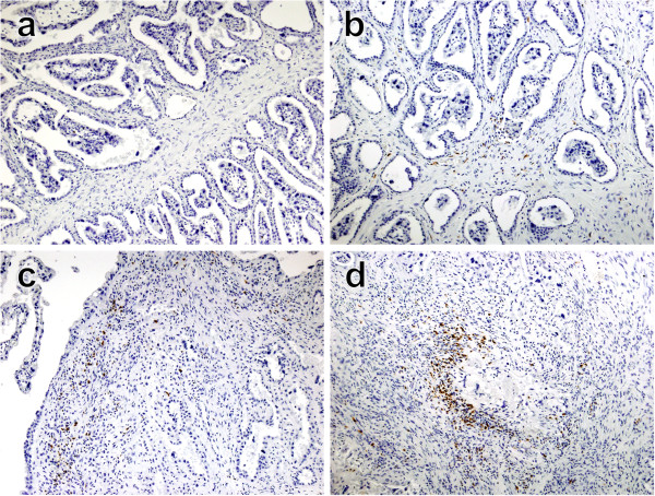
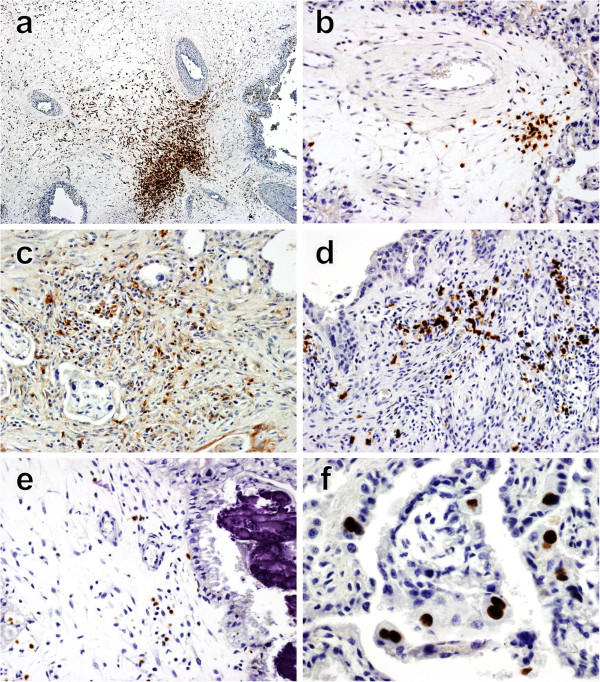
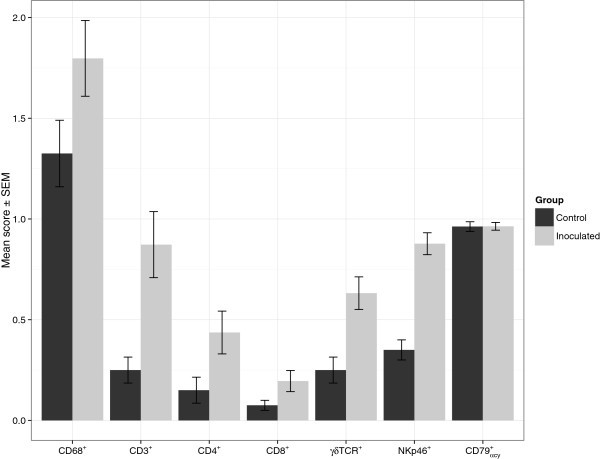
Despite Neospora caninum being a major cause of bovine abortion worldwide, its pathogenesis is not completely understood. Neospora infection stimulates host cell-mediated immune responses, which may be responsible for the placental damage leading to abortion. The aim of the current study was to characterize the placental immune response following an experimental inoculation of pregnant cattle with N. caninum tachyzoites at day 210 of gestation. Cows were culled at 14, 28, 42 and 56 days post inoculation (dpi). Placentomes were examined by immunohistochemistry using antibodies against macrophages, T-cell subsets (CD4, CD8 and γδ), NK cells and B cells. Macrophages were detected mainly at 14 days post inoculation. Inflammation was generally mild and mainly characterized by CD3+, CD4+ and γδ T-cells; whereas CD8+ and NK cells were less numerous. The immune cell repertoire observed in this study was similar to those seen in pregnant cattle challenged with N. caninum at early gestation. However, cellular infiltrates were less severe than those seen during first trimester Neospora infections. This may explain the milder clinical outcome observed when animals are infected late in gestation.
Figures
Similar articles
-
Inflammatory infiltration into placentas of Neospora caninum challenged cattle correlates with clinical outcome of pregnancy.Vet Res. 2014 Jan 31;45(1):11. doi: 10.1186/1297-9716-45-11. Vet Res. 2014. PMID: 24484200 Free PMC article.
-
Cytokine expression in the placenta of pregnant cattle after inoculation with Neospora caninum.Vet Immunol Immunopathol. 2014 Sep 15;161(1-2):77-89. doi: 10.1016/j.vetimm.2014.07.004. Epub 2014 Jul 18. Vet Immunol Immunopathol. 2014. PMID: 25091332
-
Characterization of the immune response in the placenta of cattle experimentally infected with Neospora caninum in early gestation.J Comp Pathol. 2006 Aug-Oct;135(2-3):130-141. doi: 10.1016/j.jcpa.2006.07.001. J Comp Pathol. 2006. PMID: 16997005
-
Immune response in bovine neosporosis: Protection or contribution to the pathogenesis of abortion.Microb Pathog. 2017 Aug;109:177-182. doi: 10.1016/j.micpath.2017.05.042. Epub 2017 May 31. Microb Pathog. 2017. PMID: 28578088 Review.
-
The host-parasite relationship in bovine neosporosis.Vet Immunol Immunopathol. 2005 Oct 18;108(1-2):29-36. doi: 10.1016/j.vetimm.2005.07.004. Vet Immunol Immunopathol. 2005. PMID: 16098610 Review.
Cited by
-
Histopathological changes in the pancreas of cattle with abdominal fat necrosis.J Vet Med Sci. 2017 Jan 20;79(1):52-59. doi: 10.1292/jvms.16-0282. Epub 2016 Oct 30. J Vet Med Sci. 2017. PMID: 27795463 Free PMC article.
-
Systemic and local immune responses in sheep after Neospora caninum experimental infection at early, mid and late gestation.Vet Res. 2016 Jan 6;47:2. doi: 10.1186/s13567-015-0290-0. Vet Res. 2016. PMID: 26739099 Free PMC article.
-
Bovine Polymorphonuclear Neutrophils Cast Neutrophil Extracellular Traps against the Abortive Parasite Neospora caninum.Front Immunol. 2017 May 29;8:606. doi: 10.3389/fimmu.2017.00606. eCollection 2017. Front Immunol. 2017. PMID: 28611772 Free PMC article.
-
Development of maternal and foetal immune responses in cattle following experimental challenge with Neospora caninum at day 210 of gestation.Vet Res. 2013 Oct 3;44(1):91. doi: 10.1186/1297-9716-44-91. Vet Res. 2013. PMID: 24090114 Free PMC article.
-
Inflammatory infiltration into placentas of Neospora caninum challenged cattle correlates with clinical outcome of pregnancy.Vet Res. 2014 Jan 31;45(1):11. doi: 10.1186/1297-9716-45-11. Vet Res. 2014. PMID: 24484200 Free PMC article.
References
-
- Anderson ML, Andrianarivo AG, Conrad PA. Neosporosis in cattle. Anim Reprod Sci. 2000;60–61:417–431. - PubMed
Publication types
MeSH terms
LinkOut - more resources
Full Text Sources
Other Literature Sources
Research Materials
