

Clinical review: Continuous and simplified electroencephalography to monitor brain recovery after cardiac arrest
- PMID: 23876221
- PMCID: PMC4056658
- DOI: 10.1186/cc12699
Clinical review: Continuous and simplified electroencephalography to monitor brain recovery after cardiac arrest
Abstract
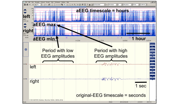
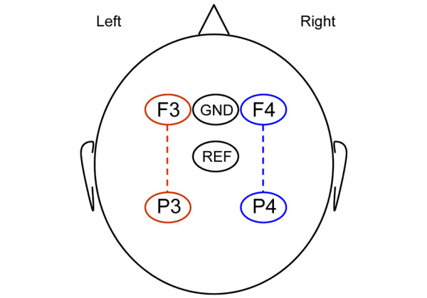
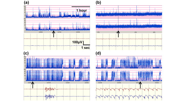
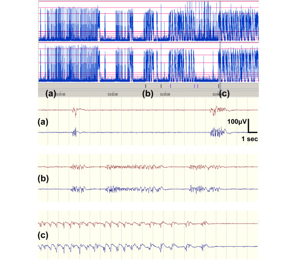
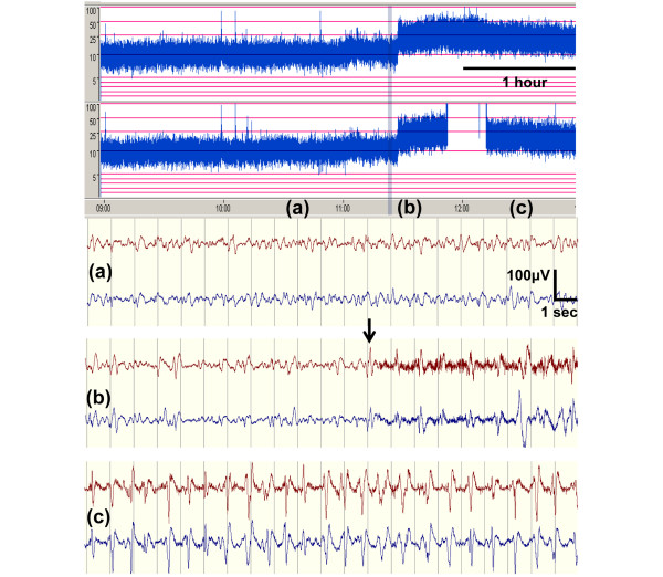
There has been a dramatic change in hospital care of cardiac arrest survivors in recent years, including the use of target temperature management (hypothermia). Clinical signs of recovery or deterioration, which previously could be observed, are now concealed by sedation, analgesia, and muscle paralysis. Seizures are common after cardiac arrest, but few centers can offer high-quality electroencephalography (EEG) monitoring around the clock. This is due primarily to its complexity and lack of resources but also to uncertainty regarding the clinical value of monitoring EEG and of treating post-ischemic electrographic seizures. Thanks to technical advances in recent years, EEG monitoring has become more available. Large amounts of EEG data can be linked within a hospital or between neighboring hospitals for expert opinion. Continuous EEG (cEEG) monitoring provides dynamic information and can be used to assess the evolution of EEG patterns and to detect seizures. cEEG can be made more simple by reducing the number of electrodes and by adding trend analysis to the original EEG curves. In our version of simplified cEEG, we combine a reduced montage, displaying two channels of the original EEG, with amplitude-integrated EEG trend curves (aEEG). This is a convenient method to monitor cerebral function in comatose patients after cardiac arrest but has yet to be validated against the gold standard, a multichannel cEEG. We recently proposed a simplified system for interpreting EEG rhythms after cardiac arrest, defining four major EEG patterns. In this topical review, we will discuss cEEG to monitor brain function after cardiac arrest in general and how a simplified cEEG, with a reduced number of electrodes and trend analysis, may facilitate and improve care.
Figures
Similar articles
-
Bedside interpretation of simplified continuous EEG after cardiac arrest.Acta Anaesthesiol Scand. 2020 Jan;64(1):85-92. doi: 10.1111/aas.13466. Epub 2019 Oct 2. Acta Anaesthesiol Scand. 2020. PMID: 31465539
-
Prognostic value of continuous EEG monitoring during therapeutic hypothermia after cardiac arrest.Crit Care. 2010;14(5):R173. doi: 10.1186/cc9276. Epub 2010 Sep 29. Crit Care. 2010. PMID: 20920227 Free PMC article.
-
Validation of the suppression ratio from a simplified EEG montage during targeted temperature management after cardiac arrest.Resuscitation. 2020 Aug;153:1-5. doi: 10.1016/j.resuscitation.2020.05.014. Epub 2020 May 20. Resuscitation. 2020. PMID: 32445782
-
Continuous electroencephalogram monitoring in the intensive care unit.Anesth Analg. 2009 Aug;109(2):506-23. doi: 10.1213/ane.0b013e3181a9d8b5. Anesth Analg. 2009. PMID: 19608827 Review.
-
Utility and rationale for continuous EEG monitoring: a primer for the general intensivist.Crit Care. 2024 Jul 16;28(1):244. doi: 10.1186/s13054-024-04986-0. Crit Care. 2024. PMID: 39014421 Free PMC article. Review.
Cited by
-
Group-Based Trajectory Modeling of Suppression Ratio After Cardiac Arrest.Neurocrit Care. 2016 Dec;25(3):415-423. doi: 10.1007/s12028-016-0263-9. Neurocrit Care. 2016. PMID: 27033709 Free PMC article.
-
How to assess prognosis after cardiac arrest and therapeutic hypothermia.Crit Care. 2014 Jan 14;18(1):202. doi: 10.1186/cc13696. Crit Care. 2014. PMID: 24417885 Free PMC article. Review.
-
Breakthrough in cardiac arrest: reports from the 4th Paris International Conference.Ann Intensive Care. 2015 Dec;5(1):22. doi: 10.1186/s13613-015-0064-x. Epub 2015 Sep 17. Ann Intensive Care. 2015. PMID: 26380990 Free PMC article.
-
Amplitude-Integrated Electroencephalography and Brain Oxygenation for Postcardiac Arrest Patients with Targeted Temperature Management.Ther Hypothermia Temp Manag. 2019 Sep;9(3):209-215. doi: 10.1089/ther.2018.0051. Epub 2019 Aug 5. Ther Hypothermia Temp Manag. 2019. PMID: 31381485 Free PMC article.
-
The Brain after Cardiac Arrest.Semin Neurol. 2017 Feb;37(1):19-24. doi: 10.1055/s-0036-1597833. Epub 2017 Feb 1. Semin Neurol. 2017. PMID: 28147414 Free PMC article. Review.
References
-
- Neumar RW, Nolan JP, Adrie C, Aibiki M, Berg RA, Bottiger BW, Callaway C, Clark RS, Geocadin RG, Jauch EC, Kern KB, Laurent I, Longstreth WT Jr, Merchant RM, Morley P, Morrison LJ, Nadkarni V, Peberdy MA, Rivers EP, Rodriguez-Nunez A, Sellke FW, Spaulding C, Sunde K, Vanden Hoek T. Post-cardiacarrest syndrome: epidemiology, pathophysiology, treatment, and prognostication. A consensus statement from the International Liaison Committee on Resuscitation (American Heart Association, Australian and New Zealand Council on Resuscitation, European Resuscitation Council, Heart and Stroke Foundation of Canada, InterAmerican Heart Foundation, Resuscitation Council of Asia, and the Resuscitation Council of Southern Africa); the American Heart Association Emergency Cardiovascular Care Committee; the Council on Cardiovascular Surgery and Anesthesia; the Council on Cardiopulmonary, Perioperative, and Critical Care; the Councilon Clinical Cardiology; and the Stroke Council. Circulation. 2008;17:2452–2483. doi: 10.1161/CIRCULATIONAHA.108.190652. - DOI - PubMed
-
- Cloostermans MC, van Meulen FB, Eertman CJ, Hom HW, van Putten MJ. Continuous electroencephalography monitoring for early prediction of neurological outcome in postanoxic patients after cardiac arrest: a prospective cohort study. Crit Care Med. 2012;17:2867–2875. doi: 10.1097/CCM.0b013e31825b94f0. - DOI - PubMed
Publication types
MeSH terms
LinkOut - more resources
Full Text Sources
Other Literature Sources
Medical
