Diet-induced obesity drives negative mouse vein graft wall remodeling
- PMID: 23876511
- PMCID: PMC3823755
- DOI: 10.1016/j.jvs.2013.05.033
Diet-induced obesity drives negative mouse vein graft wall remodeling
Abstract
Introduction: The heightened inflammatory phenotype associated with obesity has been linked to the development of cardiovascular diseases. Short-term high-fat feeding induces a proinflammatory state that may impact the blood vessel wall. CD11c, a significantly increased dendritic cell biomarker during diet-induced obesity (DIO), may have a mechanistic role in this high-fat feeding effect. We hypothesized that the proinflammatory effect of short-term DIO accelerates vein bypass graft failure via CD11c-dependent mechanisms.
Methods: Male 9-week-old DIO mice (n = 13, C57BL/6J recipients; n = 6, CD11c(-/-) recipients) and normal chow controls (n = 15, C57BL/6J recipients; n = 6, CD11c(-/-) recipients) underwent unilateral carotid interposition vein isografting (inferior vena cava from the same diet and genetic background donor), with a midgraft or outflow focal stenosis. Vein grafts were harvested at either 1 week (immunohistochemical staining for early CD11c expression) or 4 weeks later (morphometric analyses and CD11c evaluation).
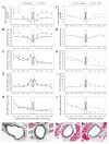
Results: Despite a 40% larger body size, C57BL/6J DIO mice had 44% smaller poststenosis vein graft lumens (P = .03) than their controls via an acceleration of overall negative vein graft wall remodeling in the day-28 midgraft focal stenosis model but not in the outflow stenosis model. Higher CD11c expression occurred in DIO midgraft-stenosis vein graft walls, both at postoperative days 7 and 28. In contrast, with in vivo CD11c deficiency, DIO did not elicit this poststenotic negative remodeling but attenuated intimal hyperplasia.
Conclusions: These findings highlight negative wall remodeling as a potential factor leading to vein graft failure and provide direct evidence that short-term dietary alterations in the mammalian metabolic milieu can have lasting implications related to acute vascular interventions. DIO induces negative mouse vein graft wall remodeling via CD11c-depedent pathways.
Copyright © 2014 Society for Vascular Surgery. Published by Mosby, Inc. All rights reserved.
Figures


Similar articles
-
Neointimal hyperplasia rapidly reaches steady state in a novel murine vein graft model.J Vasc Surg. 2002 Oct;36(4):824-32. J Vasc Surg. 2002. PMID: 12368745
-
Mouse vein graft hemodynamic manipulations to enhance experimental utility.Am J Pathol. 2011 Jun;178(6):2910-9. doi: 10.1016/j.ajpath.2011.02.014. Am J Pathol. 2011. PMID: 21641408 Free PMC article.
-
Short-term preoperative protein restriction attenuates vein graft disease via induction of cystathionine γ-lyase.Cardiovasc Res. 2020 Feb 1;116(2):416-428. doi: 10.1093/cvr/cvz086. Cardiovasc Res. 2020. PMID: 30924866 Free PMC article.
-
Perivascular innate immune events modulate early murine vein graft adaptations.J Vasc Surg. 2013 Feb;57(2):486-492.e2. doi: 10.1016/j.jvs.2012.07.007. Epub 2012 Nov 3. J Vasc Surg. 2013. PMID: 23127978 Free PMC article.
-
Targets for gene therapy of vein grafts.Curr Opin Cardiol. 1999 Nov;14(6):489-94. doi: 10.1097/00001573-199911000-00007. Curr Opin Cardiol. 1999. PMID: 10579065 Review.
Cited by
-
Role of Perivascular Adipose Tissue in Vein Remodeling.Arterioscler Thromb Vasc Biol. 2025 May;45(5):576-584. doi: 10.1161/ATVBAHA.124.321692. Epub 2025 Mar 13. Arterioscler Thromb Vasc Biol. 2025. PMID: 40079141 Free PMC article. Review.
-
A Novel Porcine Model of Bilateral Hindlimb Bypass Graft Surgery Integrating Transit Time Flowmetry.J Cardiothorac Surg. 2024 Dec 20;19(1):661. doi: 10.1186/s13019-024-03192-x. J Cardiothorac Surg. 2024. PMID: 39702209 Free PMC article.
-
Tumor necrosis factor alpha-stimulated gene-6 (TSG-6) inhibits the inflammatory response by inhibiting the activation of P38 and JNK signaling pathway and decreases the restenosis of vein grafts in rats.Heart Vessels. 2017 Dec;32(12):1536-1545. doi: 10.1007/s00380-017-1059-3. Epub 2017 Oct 3. Heart Vessels. 2017. PMID: 28975447
-
Local Adipose-Associated Mediators and Adaptations Following Arteriovenous Fistula Creation.Kidney Int Rep. 2018 Mar 2;3(4):970-978. doi: 10.1016/j.ekir.2018.02.008. eCollection 2018 Jul. Kidney Int Rep. 2018. PMID: 29988980 Free PMC article.
References
-
- Alexander JH, Hafley G, Harrington RA, Peterson ED, Ferguson TB, Jr, Lorenz TJ, et al. Efficacy and safety of edifoligide, an E2F transcription factor decoy, for prevention of vein graft failure following coronary artery bypass graft surgery: PREVENT IV: a randomized controlled trial. JAMA. 2005;294:2446–54. - PubMed
-
- Conte MS, Bandyk DF, Clowes AW, Moneta GL, Seely L, Lorenz TJ, et al. Results of PREVENT III: a multicenter, randomized trial of edifoligide for the prevention of vein graft failure in lower extremity bypass surgery. J Vasc Surg. 2006;43:742–51. discussion 51. - PubMed
-
- Zhang L, Peppel K, Brian L, Chien L, Freedman NJ. Vein graft neointimal hyperplasia is exacerbated by tumor necrosis factor receptor-1 signaling in graft-intrinsic cells. Arterioscler Thromb Vasc Biol. 2004;24:2277–83. - PubMed
-
- Sharony R, Pintucci G, Saunders PC, Grossi EA, Baumann FG, Galloway AC, et al. Matrix metalloproteinase expression in vein grafts: role of inflammatory mediators and extracellular signal-regulated kinases-1 and -2. Am J Physiol Heart Circ Physiol. 2006;290:H1651–H9. - PubMed
Publication types
MeSH terms
Substances
Grants and funding
LinkOut - more resources
Full Text Sources
Other Literature Sources
Medical
Research Materials

