

Perinatally administered bisphenol a as a potential mammary gland carcinogen in rats
- PMID: 23876597
- PMCID: PMC3764091
- DOI: 10.1289/ehp.1306734
Perinatally administered bisphenol a as a potential mammary gland carcinogen in rats
Abstract
Background: Environmental exposure to bisphenol A (BPA) affects mammary gland development in rodents and primates. Prenatal exposure to environmentally relevant doses of BPA increased the number of intraductal hyperplasias and ductal carcinomas in situ by 50 days of age in Wistar-Furth rats.
Objective: We aimed to determine whether BPA exposure of dams during gestation only or throughout lactation affects the incidence of mammary gland neoplasia in female offspring.
Methods: We treated pregnant Sprague-Dawley rats with BPA at 0, 0.25, 2.5, 25, or 250 μg BPA/kg BW/day from gestational day (GD) 9 to birth and from GD9 to postnatal day (PND) 21. Mammary glands from BPA-exposed offspring were examined at four time points for preneoplastic and neoplastic lesions. To assess circulating BPA levels, we exposed pregnant rats to vehicle or 250 μg BPA/kg BW/day during gestation only or during gestation/lactation and analyzed sera from dams, fetuses, and nursing pups for total and unconjugated BPA.
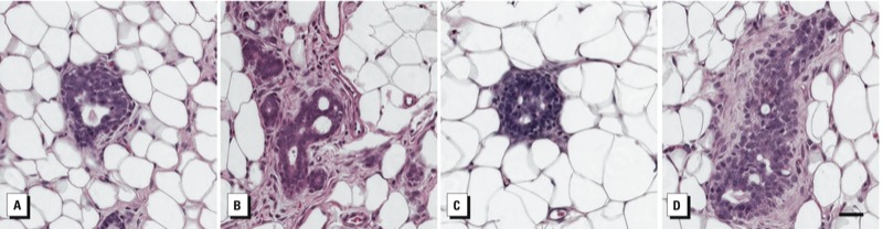
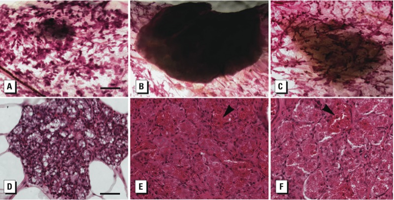
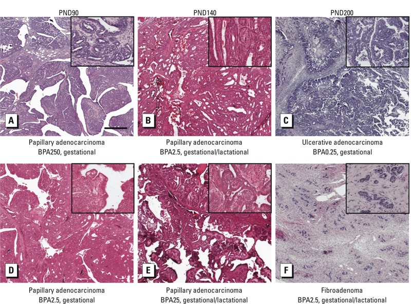
Results: Total and unconjugated BPA were detected in sera from 100% of dams and fetuses and 33% of pups exposed to 250 μg BPA/kg BW/day. Unconjugated BPA levels in exposed dams and fetuses (gestational) and in exposed dams and pups (gestational/lactational) were within levels found in humans. Preneoplastic lesions developed in BPA-exposed female offspring across all doses as early as PND50. Unexpectedly, mammary gland adenocarcinomas developed in BPA-exposed offspring by PND90.
Conclusions: Our findings suggest that developmental exposure to environmentally relevant levels of BPA during gestation and lactation induces mammary gland neoplasms in the absence of any additional carcinogenic treatment. Thus, BPA may act as a complete mammary gland carcinogen.
Conflict of interest statement
The findings and conclusions in this report are those of the authors and do not necessarily represent the views of the NIEHS or NIH.
The authors declare they have no actual or potential competing financial interests.
Figures
Comment in
-
BPA as a mammary carcinogen: early findings reported in rats.Environ Health Perspect. 2013 Sep;121(9):A284. doi: 10.1289/ehp.121-a284. Environ Health Perspect. 2013. PMID: 24004942 Free PMC article. No abstract available.
References
-
- Biegel LB, Flaws JA, Hirshfield AN, O’Connor JC, Elliot GS, Ladics GS, et al. 90-day feeding and one-generation reproduction study in Crl:CD BR rats with 17β-estradiol. Toxicol Sci. 1998;44:116–142. - PubMed
-
- Braun MM, Ahlbom A, Floderus B, Brinton LA, Hoover RN. Effect of twinship on incidence of cancer of the testis, breast, and other sites (Sweden). Cancer Causes Control. 1995;6:519–524. - PubMed
-
- Burridge E. 2008. Chemical profile: bisphenol A. ICIS Chem Business 274:48.
Publication types
MeSH terms
Substances
Grants and funding
LinkOut - more resources
Full Text Sources
Other Literature Sources