

A molecular mechanism for glaucoma: endoplasmic reticulum stress and the unfolded protein response
- PMID: 23876925
- PMCID: PMC3795998
- DOI: 10.1016/j.molmed.2013.06.005
A molecular mechanism for glaucoma: endoplasmic reticulum stress and the unfolded protein response
Abstract
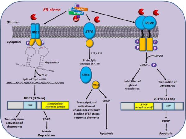
Primary open angle glaucoma (POAG) is a common late-onset neurodegenerative disease. Ocular hypertension represents a major risk factor, but POAG etiology remains poorly understood. Some cases of early-onset congenital glaucoma and adult POAG are linked to mutations in myocilin, a secreted protein of poorly defined function. Transgenic overexpression of myocilin in Drosophila and experiments in mice and human populations implicate the unfolded protein response (UPR) in the pathogenesis of glaucoma. We postulate that compromised ability of the UPR to eliminate misfolded mutant or damaged proteins, including myocilin, causes endoplasmic reticulum stress, resulting in functional impairment of trabecular meshwork cells that regulate intraocular pressure. This mechanism of POAG is reminiscent of other age-dependent neurodegenerative diseases that involve accumulation of protein aggregates.
Keywords: myocilin; neurodegenerative disease; ocular hypertension; primary open angle glaucoma.
Copyright © 2013 Elsevier Ltd. All rights reserved.
Figures
Similar articles
-
Aggregated myocilin induces russell bodies and causes apoptosis: implications for the pathogenesis of myocilin-caused primary open-angle glaucoma.Am J Pathol. 2007 Jan;170(1):100-9. doi: 10.2353/ajpath.2007.060806. Am J Pathol. 2007. PMID: 17200186 Free PMC article.
-
Overexpression of myocilin in the Drosophila eye activates the unfolded protein response: implications for glaucoma.PLoS One. 2009;4(1):e4216. doi: 10.1371/journal.pone.0004216. Epub 2009 Jan 16. PLoS One. 2009. PMID: 19148291 Free PMC article.
-
Histochemical Analysis of Glaucoma Caused by a Myocilin Mutation in a Human Donor Eye.Ophthalmol Glaucoma. 2018 Sep-Oct;1(2):132-138. doi: 10.1016/j.ogla.2018.08.004. Epub 2018 Aug 17. Ophthalmol Glaucoma. 2018. PMID: 30906929 Free PMC article.
-
Targeting the ER-autophagy system in the trabecular meshwork to treat glaucoma.Exp Eye Res. 2016 Mar;144:38-45. doi: 10.1016/j.exer.2015.08.017. Epub 2015 Aug 22. Exp Eye Res. 2016. PMID: 26302411 Free PMC article. Review.
-
The Genetic and Endoplasmic Reticulum-Mediated Molecular Mechanisms of Primary Open-Angle Glaucoma.Int J Mol Sci. 2020 Jun 11;21(11):4171. doi: 10.3390/ijms21114171. Int J Mol Sci. 2020. PMID: 32545285 Free PMC article. Review.
Cited by
-
Age at Glaucoma Diagnosis in Germline Myocilin Mutation Patients: Associations with Polymorphisms in Protein Stabilities.Genes (Basel). 2021 Nov 16;12(11):1802. doi: 10.3390/genes12111802. Genes (Basel). 2021. PMID: 34828408 Free PMC article. Review.
-
Cellular processing of myocilin.PLoS One. 2014 Apr 14;9(4):e92845. doi: 10.1371/journal.pone.0092845. eCollection 2014. PLoS One. 2014. PMID: 24732711 Free PMC article.
-
Genetics of Primary Inherited Disorders of the Optic Nerve: Clinical Applications.Cold Spring Harb Perspect Med. 2015 Jul 1;5(7):a017277. doi: 10.1101/cshperspect.a017277. Cold Spring Harb Perspect Med. 2015. PMID: 26134840 Free PMC article. Review.
-
Long-term and potent IOP-lowering effect of IκBα-siRNA in a nonhuman primate model of chronic ocular hypertension.iScience. 2022 Mar 22;25(4):104149. doi: 10.1016/j.isci.2022.104149. eCollection 2022 Apr 15. iScience. 2022. PMID: 35445186 Free PMC article.
-
Autophagy stimulation reduces ocular hypertension in a murine glaucoma model via autophagic degradation of mutant myocilin.JCI Insight. 2021 Mar 8;6(5):e143359. doi: 10.1172/jci.insight.143359. JCI Insight. 2021. PMID: 33539326 Free PMC article.
References
-
- Stone EM, et al. Identification of a gene that causes primary open angle glaucoma. Science. 1997;275:668–670. - PubMed
-
- Sheffield VC, et al. Genetic linkage of familial open angle glaucoma to chromosome 1q21-q31. Nature Genetics. 1993;4:47–50. - PubMed
-
- Michels-Rautenstrauss KG, et al. Juvenile open angle glaucoma: fine mapping of the TIGR gene to 1q24.3-q25.2 and mutation analysis. Hum. Genet. 1998;102:103–106. - PubMed
-
- Clark AF, et al. Glucocorticoid induction of the glaucoma gene MYOC in human and monkey trabecular meshwork cells and tissues. Invest. Ophthalmol. Vis. Sci. 2001;42:1769–1780. - PubMed
Publication types
MeSH terms
Substances
Grants and funding
LinkOut - more resources
Full Text Sources
Other Literature Sources
Medical
