An HPLC-tandem mass spectrometry method for simultaneous detection of alkylated base excision repair products
- PMID: 23876937
- PMCID: PMC3812247
- DOI: 10.1016/j.ymeth.2013.07.020
An HPLC-tandem mass spectrometry method for simultaneous detection of alkylated base excision repair products
Abstract
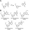
DNA glycosylases excise a broad spectrum of alkylated, oxidized, and deaminated nucleobases from DNA as the initial step in base excision repair. Substrate specificity and base excision activity are typically characterized by monitoring the release of modified nucleobases either from a genomic DNA substrate that has been treated with a modifying agent or from a synthetic oligonucleotide containing a defined lesion of interest. Detection of nucleobases from genomic DNA has traditionally involved HPLC separation and scintillation detection of radiolabeled nucleobases, which in the case of alkylation adducts can be laborious and costly. Here, we describe a mass spectrometry method to simultaneously detect and quantify multiple alkylpurine adducts released from genomic DNA that has been treated with N-methyl-N-nitrosourea (MNU). We illustrate the utility of this method by monitoring the excision of N3-methyladenine (3 mA) and N7-methylguanine (7 mG) by a panel of previously characterized prokaryotic and eukaryotic alkylpurine DNA glycosylases, enabling a comparison of substrate specificity and enzyme activity by various methods. Detailed protocols for these methods, along with preparation of genomic and oligonucleotide alkyl-DNA substrates, are also described.
Keywords: 1,N(6)-ethenoadenine; 1mA; 3mA; 4-(2-hydroxyethyl)-1-piperazineethanesulfonic acid; 6-carboxyfluorescein; 7mG; AAG; AlkA; Alkylation; BSA; Base excision repair; CID; DMS; DNA glycosylase; DTT; E. coli 3-methyladenine DNA glycosylase I; E. coli 3-methyladenine DNA glycosylase II; EDTA; ESI; ESI(+); FAM; HEPES; HPLC; MAG; MMS; MNNG; MNU; MRM; MS/MS; Mass spectrometry; Methylpurine; N-methyl-N-nitrosourea; N-methyl-N′-nitro-N-nitrosoguanidine; N1-methyladenine; N3-methyladenine; N7-methylguanine; TAG; Tris; bovine serum albumin; collision induced dissociation; dimethylsulfate; dithiothreitol; electrospray ionization; ethylenediaminetetraacetic acid; high performance liquid chromatrography; human alkyladenine DNA glycosylase; methyladenine DNA glycosylase; methylmethanesulfonate; multiple reaction monitoring; positive ion mode electrospray ionization; tandem mass spectrometry; tris(hydroxymethyl)aminomethane; εA.
Copyright © 2013 Elsevier Inc. All rights reserved.
Figures


References
-
- Friedberg E, Walker G, Siede W, Wood R, Schultz R, T E. DNA Repair and Mutagenesis. 2nd. ASM Press; Washimgton, DC: 2006.
-
- Lawley P. In: Chemical Carcinogens. Searle CE, editor. American Chemical Society; Washington, D.C.: 1976. pp. 325–484.
-
- Sedgwick B. Nat Rev Mol Cell Biol. 2004;5:148–157. - PubMed
-
- Fromme JC, Banerjee A, Verdine GL. Curr Opin Struct Biol. 2004;14:43–49. - PubMed
Publication types
MeSH terms
Substances
Grants and funding
LinkOut - more resources
Full Text Sources
Other Literature Sources
Research Materials

