. 2013 Sep;21(9):1668-77.
doi: 10.1038/mt.2013.90. Epub 2013 Jul 23.

Disruption of TAB1/p38α interaction using a cell-permeable peptide limits myocardial ischemia/reperfusion injury

Affiliations

Disruption of TAB1/p38α interaction using a cell-permeable peptide limits myocardial ischemia/reperfusion injury

Qingyang Wang et al. Mol Ther. 2013 Sep.

Abstract

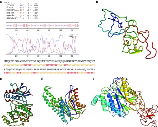
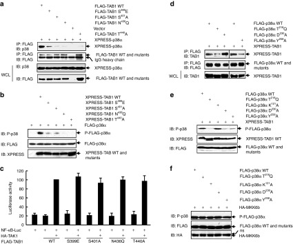
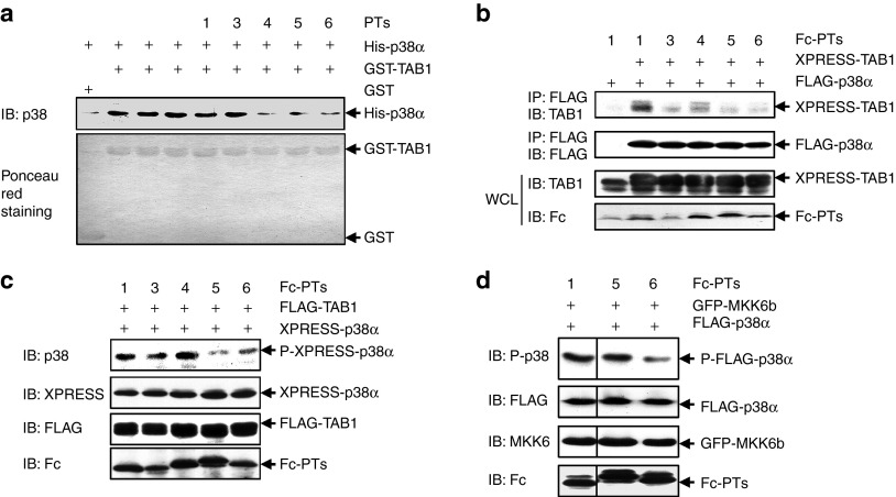
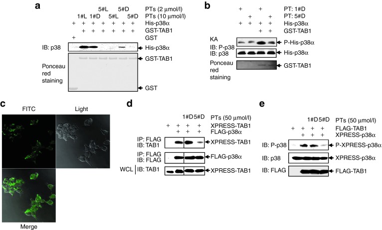
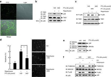
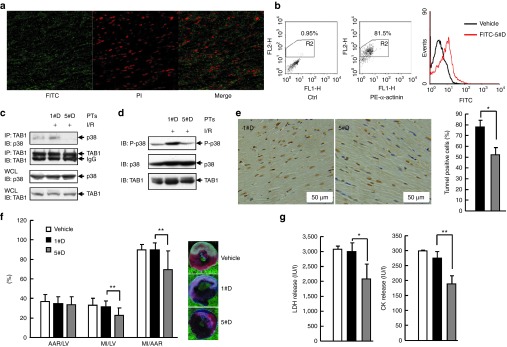
Targeting the adaptor protein (transforming growth factor-β (TGF-β)-activated protein kinase 1 (TAK1)-binding protein 1) (TAB1)-mediated non-canonical activation of p38α to limit ischemia/reperfusion (I/R) injury after an acute myocardial infarction seems to be attractive since TAB1/p38α interaction occurs specifically in very limited circumstances and possesses unique structural basis. However, so far no TAB1/p38α interaction inhibitor has been reported due to the limited knowledge about the interfaces. In this study, we sought to identify key amino acids essential for the unique mode of interaction with computer-guided molecular simulations and molecular docking. After validation of the predicted three-dimensional (3-D) structure of TAB1/p38α complex, we designed several peptides and evaluated whether they could block TAB1/p38α interaction with selectivity. We found that a cell-permeable peptide worked as a selective TAB1/p38α interaction inhibitor and decreased myocardial I/R injury. To our knowledge, this is the first TAB1/p38α interaction inhibitor.


Figures

Figure 1
Figure 1
Molecular modeling of the three-dimensional (3-D) ribbon structures of human p38α and human TAB1 in the solvent. (a) The predicted secondary structure of TAB1ΔN with GOR IV method. (b) The optimized 3-D structure of TAB1ΔN based on ab initio modeling method. (c,d) The optimized 3-D structures of (c) p38α and (d) TAB1ΔC under CVFF and Charmm force field. (e) The optimized 3-D structure of full length TAB1 under CVFF and AMBER force field.
Figure 2
Figure 2
The three-dimensional (3-D) modeling structure of TAB1/p38α complex. (a,b) The calculated surface electrostatic potential distribution of (a) p38α and (b) TAB1 based on DELPHI program. The red, blue, and white regions denote the negative electrostatic potential, the positive electrostatic potential, and the neutral electrostatic potential, respectively. (ce) The predicted 3-D structure of TAB1/p38α complex (as shown in c). In this model, both (d) hydrophobic interaction and (e) electrostatic effect are involved in the binding. The red line denotes the main-chain carbon atom orientation of TAB1, and the green line denotes that of p38α. (f) The predicated key sites in the unique mode of TAB1/p38α interaction. The red line denotes the main-chain carbon atom orientation of TAB1, and the purple line denotes that of p38α.
Figure 3
Figure 3
Ser399, Ser401, Asn436, and Thr440 in TAB1 as well as Thr218, Asp230, and Tyr258 in p38α are critical to the unique mode of interaction. (a,b) 293T cells were cotransfected with tagged-p38α and tagged-TAB1 wild-type (WT) or TAB1 mutants. (a) Immunoprecipitation (IP) of cell lysates with an anti-FLAG monoclonal antibody was followed by immunoblotting (IB) with an anti-p38 antibody. Densitometric readings were shown in Supplementary Figure S3a. (b) How the mutation might affect TAB1 co-overexpression–dependent p38α phosphorylation was analyzed by IB. Densitometric readings were shown in Supplementary Figure S3b. (c) 293T cells were cotransfected with the nuclear factor-κB (NF-κB) reporter pNF-κB-Luc, the control reporter pRL-TK, HA-TAK1, and FLAG-tagged TAB1 WT or TAB1 mutants. Luciferase activity was measured 36 hours after transfection. (d,e) 293T cells were cotransfected with XPRESS-TAB1 and FLAG-tagged p38α WT or p38α mutants. (d) IP of cell lysates with an anti-FLAG monoclonal antibody was followed by IB with an anti-TAB1 antibody. Densitometric readings were shown in Supplementary Figure S3c. (e) How the mutation might affect TAB1 co-overexpression–dependent p38α phosphorylation was analyzed by IB. Densitometric readings were shown in Supplementary Figure S3d. (f) 293T cells were cotransfected with HA-MKK6b and FLAG-tagged p38α WT or p38α mutants. The phosphorylation of FLAG-p38α and expression of HA-MKK6b and FLAG-p38α were analyzed by IB. Densitometric readings were shown in Supplementary Figure S3e. ns, not specific.
Figure 4
Figure 4
Design and validation of candidate peptides potentially blocking the interaction. (a) Glutathione-Sepharose beads bound with Escherichia coli (E. coli)-derived GST-TAB1 or GST were incubated with E. coli-derived His-p38α in the presence or absence of chemically synthesized peptides (final concentration: 2 μmol/l). After washing the beads, the bound proteins were eluted and subjected to SDS-PAGE and subsequent immunoblotting (IB) analysis. Densitometric readings were shown in Supplementary Figure S4a. (b,c) 293T cells were cotransfected with tagged-TAB1, tagged-p38α, and a vector encoding various peptides in fusion with the Fc portion of a human IgG1. (b) Immunoprecipitation of cell lysates with an anti-FLAG monoclonal antibody was followed by IB with an anti-TAB1 antibody. Densitometric readings were shown in Supplementary Figure S4b. (c) How the peptides might affect TAB1 co-overexpression–dependent p38α phosphorylation was analyzed by IB. Densitometric readings were shown in Supplementary Figure S4c. (d) 293T cells were cotransfected with GFP-MKK6b, FLAG-p38α, and a vector encoding various peptides in fusion with the Fc portion of a human IgG1. How the peptides might affect MKK6 co-overexpression–dependent p38 phosphorylation was analyzed by IB. Densitometric readings were shown in Supplementary Figure S4d. GFP, green fluorescent protein; GST, glutathione S-transferase; SDS-PAGE, sodium dodecyl sulfate polyacrylamide gel electrophoresis.
Figure 5
Figure 5
The cell-penetrating and protease-resistant form of peptide PT5 (5#D-PT) blocks co-overexpression–induced TAB1/p38α interaction. (a) GST-pull down assays were performed as described in Figure 4a. Densitometric readings were shown in Supplementary Figure S5a. (b) Escherichia coli (E. coli)-derived His-p38α was incubated with or without E. coli-derived GST-TAB1 in the presence of 1#D-PT or 5#D-PT for 60 minutes at 30 °C in kinase buffer. A total of 20 μmol/l nonradioactive adenosine triphosphate was included to drive the kinase reaction. The samples were then subjected to SDS-PAGE and immunoblotting (IB) with the indicated antibodies. Densitometric readings were shown in Supplementary Figure S5b. (c) 293T cells were incubated with 50 μmol/l FITC-conjugated 5#D-PT for 30 minutes. The images were captured by confocal microscopy. (d,e) 293T cells were cotransfected with tagged-TAB1 and tagged-p38α. 1#D-PT or 5#D-PT was added into the medium 6 hours later. (d) Cell lysates were harvested 30 hours post-transfection. Immunoprecipitation of cell lysates with an anti-FLAG monoclonal antibody was followed by IB with an anti-TAB1 antibody. Densitometric readings were shown in Supplementary Figure S5c. (e) How 5#D-PT might affect TAB1 co-overexpression–dependent p38α phosphorylation was analyzed by IB. Densitometric readings were shown in Supplementary Figure S5d. FITC, fluorescein isothiocyanate; GST, glutathione S-transferase; KA, kinase assay; SDS-PAGE, sodium dodecyl sulfate polyacrylamide gel electrophoresis.
Figure 6
Figure 6
5#D-PT blocks the interaction of endogenous TAB1 and endogenous p38α during simulated I/R with selectivity. (a) Rat cardiomyocytes were incubated with 50 μmol/l FITC-conjugated 5#D-PT for 30 minutes. The images were captured by confocal microscopy. (b) Cardiomyocytes were cultured under normoxic conditions or exposed to 60 minutes of simulated ischemia followed by 10 minutes of reperfusion in the presence of 1#D-PT or 5#D-PT. After cell lysates were harvested, anti-TAB1 immunoprecipitation followed by anti-p38 MAPK immunoblotting was performed. Densitometric readings were shown in Supplementary Figure S6a. (c) Cardiomyocytes were treated as described as in b. After cell lysates were harvested, how 5#D-PT might affect endogenous TAB1-mediated endogenous p38α phosphorylation was analyzed by immunoblotting (IB). Densitometric readings were shown in Supplementary Figure S6b. (d) Cardiomyocytes were cultured under normoxic conditions or exposed to 60 minutes of simulated ischemia followed by 60 minutes of reperfusion in the presence of 1#D-PT or 5#D-PT. Apoptosis was quantified by Hoechst staining. Left, mean ± SD (n = 3), *P < 0.05; right, representative images. (e) Cardiomyocytes were cultured in the presence of 1#D-PT or 5#D-PT for 60 minutes. After cell lysates were harvested, anti-MKK3b immunoprecipitation followed by anti-p38 MAPK IB was performed. Densitometric readings were shown in Supplementary Figure S6c. (f) Cardiomyocytes were stimulated with or without 1 μg/ml anisomycin or 60 J/m2 ultraviolet and incubated for 20 minutes. Phosphorylation and expression of endogenous MKK3/6 and endogenous p38 were analyzed by IB. Densitometric readings were shown in Supplementary Figure S6d. FITC, fluorescein isothiocyanate; I/R, ischemia/reperfusion.
Figure 7
Figure 7
5#D-PT reduces myocardial I/R injury in rats. (a,b) FITC-conjugated 5#D-PT (green) was administered by a single intravenous bolus injection 30 minutes before reperfusion. (a) Heart tissue was harvested 30 minutes after reperfusion and was cut in a cryomicrotome (−20 °C). After counterstained with propidium iodide (PI, red), tissue sections were subjected to confocal microscopy. (b) Adult primary cardiomyocytes were isolated 30 minutes after reperfusion and were subjected to intracellular staining with an antibody against α-actinin followed by flow cytometry analysis. α-Actinin+ cells were gated and histograms of FITC intensity were shown. (c) Rats were subjected to myocardial I/R or left untreated, proteins were isolated from heart tissue frozen 30 minutes after reperfusion. Then, anti-TAB1 immunoprecipitation followed by anti-p38 MAPK immunoblotting (IB) was performed. Densitometric readings were shown in Supplementary Figure S7a. (d) Rats were treated as described as in c. After cell lysates were harvested, how 5#D-PT might affect endogenous TAB1-mediated endogenous p38 phosphorylation in vivo was analyzed by IB. Densitometric readings were shown in Supplementary Figure S7b. (e) Apoptotic cardiomyocytes in the infarct border zone 24 hours after myocardial I/R was quantified by terminal deoxynucleotidyl transferase-mediated dUTP nick end-labeling (TUNEL) assay and hematoxylin staining. Left, representative images; right, mean ± SD. *P < 0.05. (f) Twenty-four hours after myocardial I/R, the left ventricle (LV) were dissected, then the area at risk (AAR) and myocardial infarct (MI) size were determined by Evans blue and 1% 2,3,5-triphenyl-tetrazolium chloride staining. Data from 9–16 mice per group are summarized. **P < 0.01. (g) Twenty-four hours after myocardial I/R, the LDH/CK levels in animal blood samples were determined. Data from 9–16 mice per group are summarized. *P < 0.05; **P < 0.01. CK, creatine kinase; FITC, fluorescein isothiocyanate; I/R, ischemia/reperfusion; LDH, lactate dehydrogenase.

References

    1. Hausenloy DJ, Yellon DM. Myocardial ischemia-reperfusion injury: a neglected therapeutic target. J Clin Invest. 2013;123:92–100. - PMC - PubMed
    1. Eltzschig HK, Eckle T. Ischemia and reperfusion–from mechanism to translation. Nat Med. 2011;17:1391–1401. - PMC - PubMed
    1. Denise Martin E, De Nicola GF, Marber MS. New therapeutic targets in cardiology: p38 alpha mitogen-activated protein kinase for ischemic heart disease. Circulation. 2012;126:357–368. - PubMed
    1. Marber MS, Rose B, Wang Y. The p38 mitogen-activated protein kinase pathway–a potential target for intervention in infarction, hypertrophy, and heart failure. J Mol Cell Cardiol. 2011;51:485–490. - PMC - PubMed
    1. Kumphune S, Bassi R, Jacquet S, Sicard P, Clark JE, Verma S, et al. A chemical genetic approach reveals that p38alpha MAPK activation by diphosphorylation aggravates myocardial infarction and is prevented by the direct binding of SB203580. J Biol Chem. 2010;285:2968–2975. - PMC - PubMed

Publication types

MeSH terms

Substances