

From AR to c-Met: androgen deprivation leads to a signaling pathway switch in prostate cancer cells
- PMID: 23877345
- PMCID: PMC3829778
- DOI: 10.3892/ijo.2013.2020
From AR to c-Met: androgen deprivation leads to a signaling pathway switch in prostate cancer cells
Abstract
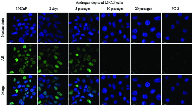
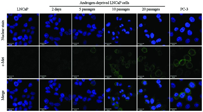
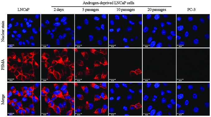
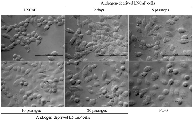
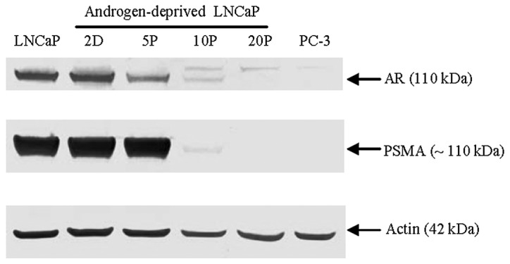
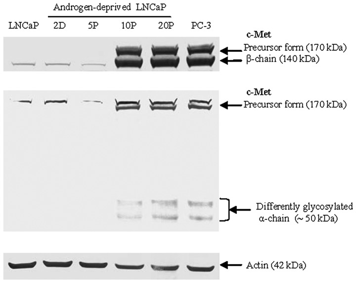
Elucidating the role of androgen deprivation in the transition from androgen-dependence to independence may enable the development of more specific therapeutic strategies against prostate cancer. Our previous in vitro model was employed to further assess the effects of continuous androgen‑deprivation on prostate cancer cells (LNCaP) with respect to both androgen receptor (AR) and c-Met expression. The results indicated that long-term androgen deprivation resulted in a signaling pathway switch from AR to c-Met in androgen-sensitive cells, which was confirmed by immunofluorescence imaging and western blot analysis. This signaling pathway switch may be predictive of a more aggressive disease state following androgen deprivation therapy.
Figures
Similar articles
-
Increased Akt signaling resulting from the loss of androgen responsiveness in prostate cancer.Curr Med Chem. 2013;20(1):144-57. Curr Med Chem. 2013. PMID: 23033951
-
Prolonged androgen deprivation leads to downregulation of androgen receptor and prostate-specific membrane antigen in prostate cancer cells.Int J Oncol. 2012 Dec;41(6):2087-92. doi: 10.3892/ijo.2012.1649. Epub 2012 Oct 4. Int J Oncol. 2012. PMID: 23041906 Free PMC article.
-
The androgen receptor negatively regulates the expression of c-Met: implications for a novel mechanism of prostate cancer progression.Cancer Res. 2007 Feb 1;67(3):967-75. doi: 10.1158/0008-5472.CAN-06-3552. Cancer Res. 2007. PMID: 17283128
-
Androgen signal transduction and prostatic carcinoma.World J Urol. 1994;12(2):99-103. doi: 10.1007/BF00184245. World J Urol. 1994. PMID: 8087144 Review.
-
Androgen action in the prostate gland.Minerva Urol Nefrol. 2012 Mar;64(1):35-49. Minerva Urol Nefrol. 2012. PMID: 22402316 Review.
Cited by
-
Met in urological cancers.Cancers (Basel). 2014 Dec 16;6(4):2387-403. doi: 10.3390/cancers6042387. Cancers (Basel). 2014. PMID: 25521854 Free PMC article. Review.
-
Androgen deprivation induces double-null prostate cancer via aberrant nuclear export and ribosomal biogenesis through HGF and Wnt activation.Nat Commun. 2024 Feb 9;15(1):1231. doi: 10.1038/s41467-024-45489-4. Nat Commun. 2024. PMID: 38336745 Free PMC article.
-
Evodiamine as the Active Compound of Evodiae fructus to Inhibit Proliferation and Migration of Prostate Cancer through PI3K/AKT/NF-κB Signaling Pathway.Dis Markers. 2022 Jul 18;2022:4399334. doi: 10.1155/2022/4399334. eCollection 2022. Dis Markers. 2022. PMID: 35899176 Free PMC article.
-
Prolonged androgen deprivation leads to overexpression of calpain 2: implications for prostate cancer progression.Int J Oncol. 2014 Feb;44(2):467-72. doi: 10.3892/ijo.2013.2196. Epub 2013 Nov 29. Int J Oncol. 2014. PMID: 24297527 Free PMC article.
-
Changes in plasma biomarkers following treatment with cabozantinib in metastatic castration-resistant prostate cancer: a post hoc analysis of an extension cohort of a phase II trial.J Transl Med. 2016 Jan 13;14:12. doi: 10.1186/s12967-015-0747-y. J Transl Med. 2016. PMID: 26762579 Free PMC article. Clinical Trial.
References
-
- Gustavsson H , Welen K , Damber JE . Transition of an androgen-dependent human prostate cancer cell line into an androgen-independent subline is associated with increased angiogenesis . Prostate . 2005 ; 62 : 364 – 373 . - PubMed
-
- Lee SO , Dutt SS , Nadiminty N , Pinder E , Liao H , Gao AC . Development of an androgen-deprivation induced and androgen suppressed human prostate cancer cell line . Prostate . 2007 ; 67 : 1293 – 1300 . - PubMed
-
- Saraon P , Jarvi K , Diamandis EP . Molecular alterations during progression of prostate cancer to androgen independence . Clin Chem . 2011 ; 57 : 1366 – 1375 . - PubMed
-
- Devlin HL , Mudryj M . Progression of prostate cancer: multiple pathways to androgen independence . Cancer Lett . 2009 ; 274 : 177 – 186 . - PubMed
Publication types
MeSH terms
Substances
Grants and funding
LinkOut - more resources
Full Text Sources
Other Literature Sources
Medical
Research Materials
Miscellaneous
