. 2013 Oct;43(4):1011-8.
doi: 10.3892/ijo.2013.2021. Epub 2013 Jul 18.

Plasma endothelial protein C receptor influences innate immune response in ovarian cancer by decreasing the population of natural killer and TH17 helper cells

Affiliations

Plasma endothelial protein C receptor influences innate immune response in ovarian cancer by decreasing the population of natural killer and TH17 helper cells

Dalel Azzazene et al. Int J Oncol. 2013 Oct.

Abstract

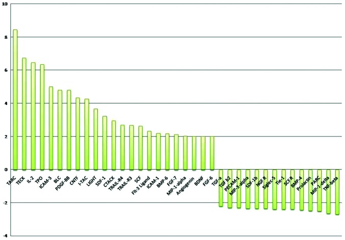
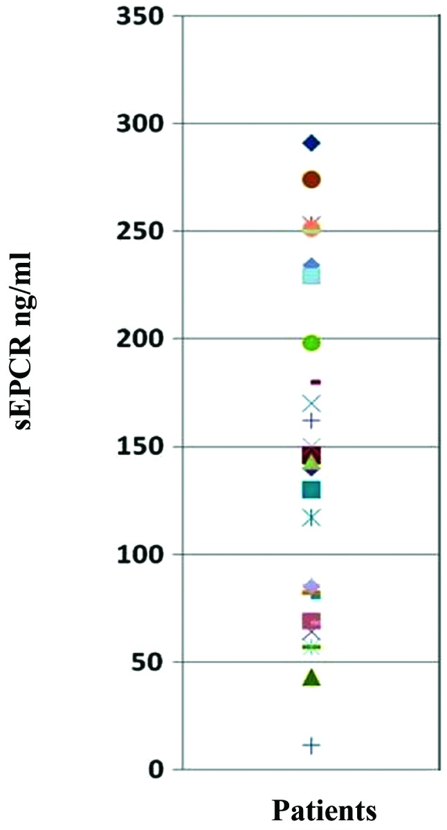
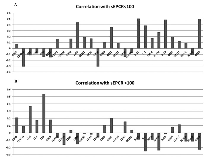
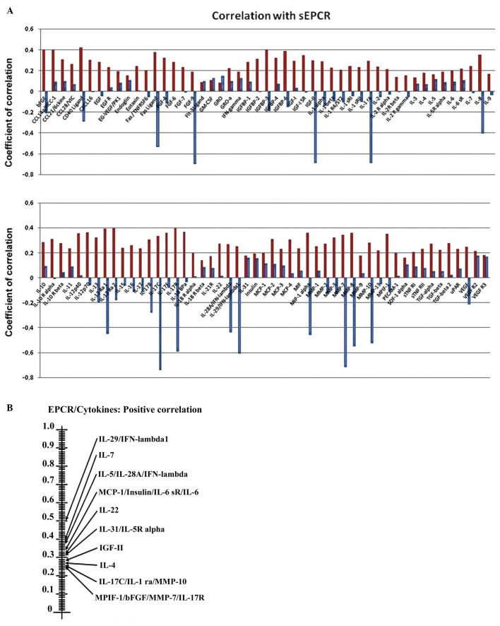
In spite of the growing importance of endothelial protein C receptor/active protein C (EPCR/aPC) in tumor biology, their impact on immunological homeostasis remains largely unexplored. The objective of this study was to assess whether soluble plasma endothelial protein C receptor (sEPCR), which is a regulator of circulating aPC, is involved in innate immune response in cancer patients. In the Ovcar-3 ovarian cancer line, the role of aPC in secretion of cytokines was analyzed. In parallel, in 33 patients, with a diagnosis of ovarian epithelial cancer, sEPCR was quantified, blood immune cell phenotypes were determined by flow cytometry and plasma cytokines were evaluated using a protein array. Spearman's rank correlation coefficients (r) and coefficient significance was determined by a statistical hypothesis test (α=0.05). Our results show that i) aPC induced the secretion of several cytokines in Ovcar-3 cells; ii) 61% of patients exhibited a concentration of plasma sEPCR well above the baseline (normal plasma level, 100 ± 28 ng/ml); iii) comparing immune cell phenotypes in patients having a normal level of sEPCR with those having a high level of sEPCR, it was found that sEPCR levels were correlated with high intensity of cells expressing CD45ra, CD3, CD8, CD25 and low intensity of cells expressing CD56 (NK cells), CD294 (TH2 cells), IL-2, IL-10, IL-17a (TH17 cells), IL-21 (TH21 cells) and CD29 markers (r ≥ 0.60); and iv) high levels of sEPCR correlate with high levels of plasma bioactive proteins such as insulin-like growth factor-2 (IGFII), IL-13rα, macrophage inflammatory protein (MIP1α) and matrix metalloproteinase-7 (MMP-7) that have already been proposed as biomarkers for ovarian cancer and particularly those with poor prognosis. In conclusion, sEPCR produced by ovarian cancer cells, by modulating circulating aPC, influences the secretory behavior of tumor cells (cytokines and interleukins). Consequently, sEPCR in turn acts on the innate immune response by decreasing effector cells such as natural killer and T helper cells (TH2, TH17 and TH21).


Figures

Figure 1.
Figure 1.
The effect of aPC on the ovarian cancer cell line. In order to analyze the pleiotropic role of active protein C (after treatments of ovarian cell line with aPC for 24 h), we examined the cell supernatants using a protein cytokine array as described in Materials and methods (RayBio ® Human Cytokine Antibody). The ratio of values between cells treated by aPC and non-treated is shown. The positive control was considered as 1. Of the 174 proteins proposed in this test only 23 were upregulated (ratio of cells treated by aPC vs. non-treated cells was >2). They are TARC, TEK, IL-2, TPO, ICAM-3, BLC, PDGF-BB, CNTF, I-TAC, LIGHT, SDF-1α, CTACK, TRAIL-R3, SCF, FLT-3 ligand, ICAM-1, BMP-6, FGF-7, MIP-1α, angiogenin, BDNF and FGF-6 while 14 proteins such as TGFα, TGFβ, PECAM-1, MIP-3α, SDF-1β, NGF-R, Siglec-5, BMP-4, prolactin, PARC, MIP-1δ and TNFβ were downregulated (ratio cells treated by aPC vs. non-treated cells was <2). The ratio situated beyond 4 was taken into consideration for the present study. The results indicate that aPC interacted with tumor cells and activated the secretion of several cytokines such as thymus and activation-regulated chemokine (TARC, CCL17), thymus expressed chemokine (TECK, CCL25), interleukin-2 (IL-2), thrombopoietin (TPO), intercellular adhesion molecule-3 (ICAM-3, CD56), B lymphocyte chemoattractant (BLC), platelet-derived growth factor-BB (PDGF-BB), ciliary neurotrophic factor (CNTF) and T cell alpha chemoattractant (I-TAC) by ovarian cell lines.
Figure 2.
Figure 2.
Evaluation of sEPCR in plasma of ovarian cancer patients. Blood (n=33) samples from patients aged >18 years were obtained. Soluble EPCR in plasma fluids was measured using Asserachrom sEPCR immunoassay as recommended by the commercial supplier. All the 33 samples tested were positive for the presence of sEPCR. The base-line value of 100±28 ng/ml was considered as normal. Thus, 60.7% of them exhibited a concentration well about the base-line.
Figure 3.
Figure 3.
Influence of plasma sEPCR in blood immune cells. Mononuclear cells from 33 patients were separated by Ficoll gradient centrifugation and then stored in 200 μ l of DMSO/FCS 50% at −80°C. Phenotyping of immuno-inflammatory cells were performed by flow cytometry the using 30 antibodies as given above. (A) When sEPCR is <100 ng/ml (group 1), plasmatic sEPCR correlated with low intensity of some cell associated markers such as CD45ra, CD3, CD8 and high intensity of with CD56 (NK cells) and cell associated IL-2, IL-10, IL-17a, IL-21 and CD29 markers (r ≤0.40). When sEPCR is >100 ng/ml (group 2), plasmatic sEPCR correlated with high intensity of CD45ra, CD3, CD8, CD25 and low intensity of CD56, IL-2, IL-10, IL-21 and CD29 markers (r ≥0.60). The expression of some cell markers such as CD4, FoxP3, CD20 and CD10 was independent of plasma sEPCR variation (r ≤0.3). A decrease in IL-17a in group-2 (B) was associated with the diminition of CD161 and ROR-γ associated cells. These results indicate that increase of plasma sEPCR was associated with a decrease of effector immune cells in blood circulation.
Figure 4.
Figure 4.
Influence sEPCR on the level of cytokines and interleukins in plasma. (A) We analyzed the correlation between the amount of sEPCR in plasma and a set of 100 biologically active plasma proteins. The membranes were incubated with serum from ovarian cancer patients (10X in PBS albumin 1%). Signal intensities were quantified with a Bio-Imaging System MF-ChemiBis 4.2 and analyzed with Multi Gauge V3.2 software. We compared the significance (r ≥0.60) of plasmatic sEPCR in group 1 (sEPCR <100, blue histograms) and in group 2 (sEPCR >100, red histograms). When plasma sEPCR is >100 ng/ml, the expression of Fas-Ligand, CD 40 ligand, bFGF, FGF-9, IGF-II, IGFbp 3 and 6, IL-ra (receptor antagonist), IL-8, IL-13ra, IL-17b, IL-17c, IL-17R, IL-28A, IL-29, MIP-1, MMP-7, MMP-9, MMP-10 (r ≥0.60) and VEGF (r= 0.40) not only reversed but also increased significantly. (B) The experimental data were further subjected to scrutiny using the Sprearman statistical test. These results confirmed the results shown in (A) and also revealed some additional ones between sEPCR levels and the recently classified type III interferon group.

References

    1. Dahlbäck B , Villoutreix BO . Molecular recognition in the protein C anticoagulant pathway . J Thromb Haemost . 2003 ; 1 : 1525 – 1534 . - PubMed
    1. Li W , Zheng X , Gu J , Ferrell GL , Lupu F , Esmon NL , Esmon CT . Overexpressing endothelial cell protein C receptor alters the hemostatic balance and protects mice from endotoxin . J Thromb Haemost . 2005 ; 3 : 1351 – 1359 . - PubMed
    1. Taylor FB , Jr , Peer GT , Lockhart MS , Ferrell G , Esmon CT . Endothelial cell protein C receptor plays an important role in protein C activation in vivo . Blood . 2001 ; 97 : 1685 – 1688 . - PubMed
    1. Fukudome K , Kurosawa S , Stearns-Kurosawa DJ , He X , Rezaie AR , Esmon CT . The endothelial cell protein C receptor. Cell surface expression and direct ligand binding by the soluble receptor . J Biol Chem . 1996 ; 271 : 17491 – 17498 . - PubMed
    1. Xu J , Qu D , Esmon NL , Esmon CT . Metalloproteolytic release of endothelial cell protein C receptor . J Biol Chem . 2000 ; 275 : 6038 – 6044 . - PubMed

MeSH terms