. 2013 Oct;57(10):4848-55.
doi: 10.1128/AAC.00762-13. Epub 2013 Jul 22.

Structures of the class D Carbapenemases OXA-23 and OXA-146: mechanistic basis of activity against carbapenems, extended-spectrum cephalosporins, and aztreonam

Affiliations

Structures of the class D Carbapenemases OXA-23 and OXA-146: mechanistic basis of activity against carbapenems, extended-spectrum cephalosporins, and aztreonam

Kip-Chumba J Kaitany et al. Antimicrob Agents Chemother. 2013 Oct.

Abstract

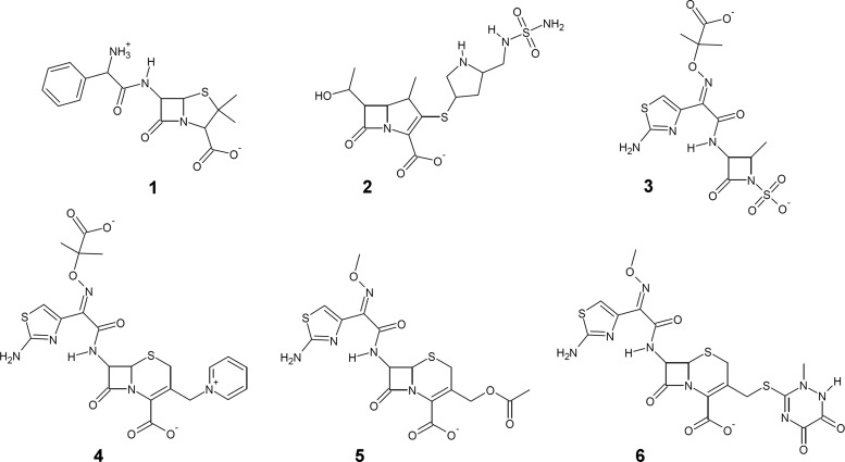
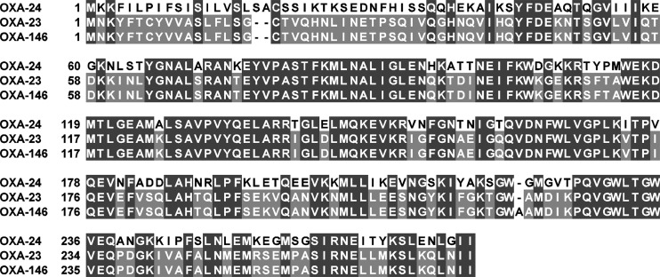
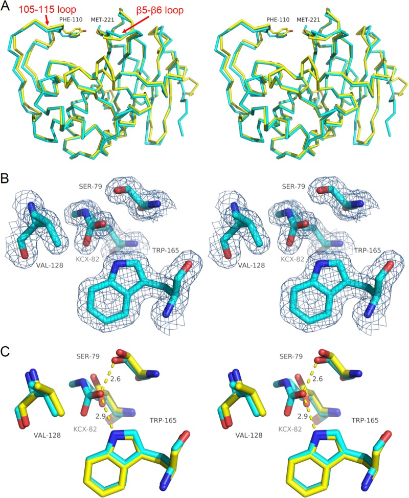
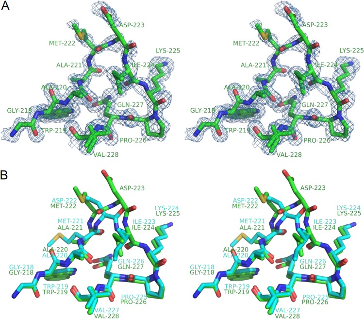
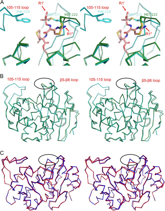
Class D β-lactamases that hydrolyze carbapenems such as imipenem and doripenem are a recognized danger to the efficacy of these "last-resort" β-lactam antibiotics. Like all known class D carbapenemases, OXA-23 cannot hydrolyze the expanded-spectrum cephalosporin ceftazidime. OXA-146 is an OXA-23 subfamily clinical variant that differs from the parent enzyme by a single alanine (A220) inserted in the loop connecting β-strands β5 and β6. We discovered that this insertion enables OXA-146 to bind and hydrolyze ceftazidime with an efficiency comparable to those of other extended-spectrum class D β-lactamases. OXA-146 also binds and hydrolyzes aztreonam, cefotaxime, ceftriaxone, and ampicillin with higher efficiency than OXA-23 and preserves activity against doripenem. In this study, we report the X-ray crystal structures of both the OXA-23 and OXA-146 enzymes at 1.6-Å and 1.2-Å resolution. A comparison of the two structures shows that the extra alanine moves a methionine (M221) out of its normal position, where it forms a bridge over the top of the active site. This single amino acid insertion also lengthens the β5-β6 loop, moving the entire backbone of this region further away from the active site. A model of ceftazidime bound in the active site reveals that these two structural alterations are both likely to relieve steric clashes between the bulky R1 side chain of ceftazidime and OXA-23. With activity against all four classes of β-lactam antibiotics, OXA-146 represents an alarming new threat to the treatment of infections caused by Acinetobacter spp.


Figures

Fig 1
Fig 1
β-Lactam antibiotics. Structures of the penicillin ampicillin (1), the carbapenem doripenem (2), the monobactam aztreonam (3), and the cephalosporins ceftazidime (4), cefotaxime (5), and ceftriaxone (6).
Fig 2
Fig 2
Multiple-sequence alignment of OXA-24, OXA-23, and OXA-146. OXA-23 and OXA-24 share 59% identity, and OXA-146 is identical to OXA-23 except for a duplication of A220.
Fig 3
Fig 3
Comparison of OXA-23 and OXA-24 structures. (A) Structural alignment of the α-carbon traces of OXA-23 (cyan) and OXA-24 (yellow; PDB accession no. 3PAE). Also shown are residues that form the bridge over the top of the active site (F110/M221 in OXA-23 and Y112/M223 in OXA-24). (B) 2Fo-Fc electron density maps of key active-site residues of OXA-23 contoured to 1.0 σ showing full carboxylation of K82. (C) Structural alignment of OXA-23 (cyan) and OXA-24 (yellow) active-site residues showing the high degree of overlap (OXA-24 structure 3PAE contains a K84D substitution).
Fig 4
Fig 4
Structure of the loop between β strands β5 and β6. (A) Stereodiagram showing OXA-146 residues 218 to 228 with 2Fo-Fc electron density maps contoured to 1.0 σ. Residue V228 is shown in multiple conformations. (B) Structural alignment of the same loop region from OXA-146 (green) and OXA-23 (cyan).
Fig 5
Fig 5
Overlaid structures showing the β5-β6 loop structural alteration. (A) Stereodiagram of OXA-146 (green) and OXA-23 (cyan) after alignment with the β-lactam sensor protein BlaR1 with ceftazidime bound as an acyl intermediate (PDB accession no. 1XKZ). The ceftazidime serine acyl moiety is shown in salmon, and the rest of the BlaR1 protein is not shown. The labels R1′ and R1″ indicate the carboxyl group and aminothiazole of the R1 side chain, respectively. The OXA-23 bridge methionine (M221) is shown in only one of its two alternative conformations, and the equivalent residue in OXA-146 (M222) is shown in its new shifted position after the insertion of the additional alanine (A221). (B) Stereodiagram showing the position of the β5-β6 loop deviation (circled) in the context of the full structures of OXA-23 and OXA-146. (C) Stereodiagram showing an alignment of SHV-1 (red; PDB accession no. 1SHV) and SHV-2 (blue; PDB accession no. 1N9B). The position of a similar loop deviation resulting in ceftazidime activity is shown (circled).

References

    1. Poirel L, Naas T, Nordmann P. 2010. Diversity, Epidemiology, and Genetics of Class D β-lactamases. Antimicrob. Agents Chemother. 54:24–38 - PMC - PubMed
    1. Naas T, Poirel L, Nordmann P. 2008. Minor extended-spectrum β-lactamases. Clin. Microbiol. Infect. 14(Suppl 1):42–52 - PubMed
    1. Walther-Rasmussen J, Hoiby N. 2006. OXA-type carbapenemases. J. Antimicrob. Chemother. 57:373–383 - PubMed
    1. Scaife W, Young HK, Paton RH, Amyes SG. 1995. Transferable imipenem-resistance in Acinetobacter species from a clinical source. J. Antimicrob. Chemother. 36:585–586 - PubMed
    1. Donald HM, Scaife W, Amyes SG, Young HK. 2000. Sequence analysis of ARI-1, a novel OXA β-lactamase, responsible for imipenem resistance in Acinetobacter baumannii 6B92. Antimicrob. Agents Chemother. 44:196–199 - PMC - PubMed

Publication types

MeSH terms

LinkOut - more resources