. 2014 Jun;71(6):2051-8.
doi: 10.1002/mrm.24871. Epub 2013 Jul 22.

Dynamic metabolic imaging of hyperpolarized [2-(13) C]pyruvate using spiral chemical shift imaging with alternating spectral band excitation

Affiliations

Dynamic metabolic imaging of hyperpolarized [2-(13) C]pyruvate using spiral chemical shift imaging with alternating spectral band excitation

Sonal Josan et al. Magn Reson Med. 2014 Jun.

Abstract

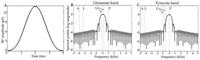
Purpose: In contrast to [1-(13) C]pyruvate, hyperpolarized [2-(13) C]pyruvate permits the ability to follow the (13) C label beyond flux through pyruvate dehydrogenase complex and investigate the incorporation of acetyl-coenzyme A into different metabolic pathways. However, chemical shift imaging (CSI) with [2-(13) C]pyruvate is challenging owing to the large spectral dispersion of the resonances, which also leads to severe chemical shift displacement artifacts for slice-selective acquisitions.

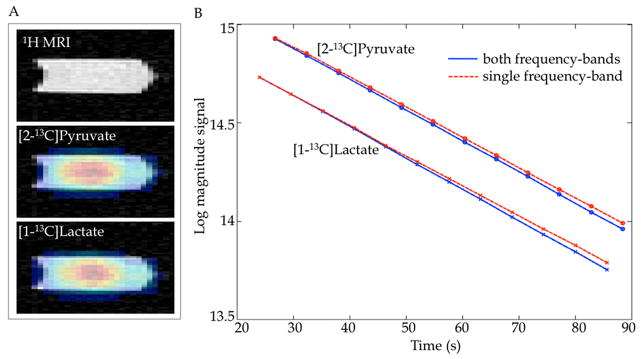
Methods: This study introduces a sequence for three-dimensional CSI of [2-(13) C]pyruvate using spectrally selective excitation of limited frequency bands containing a subset of metabolites. Dynamic CSI data were acquired alternately from multiple frequency bands in phantoms for sequence testing and in vivo in rat heart.

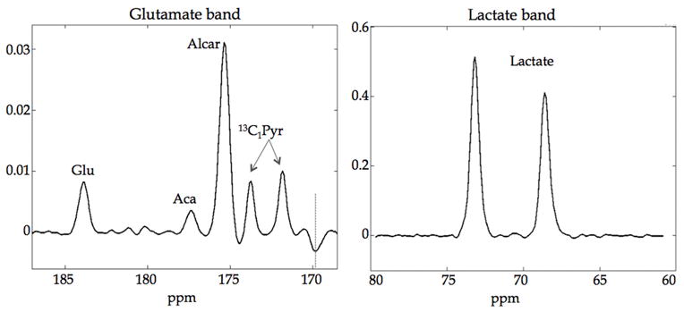
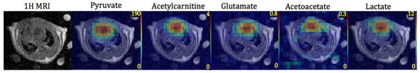
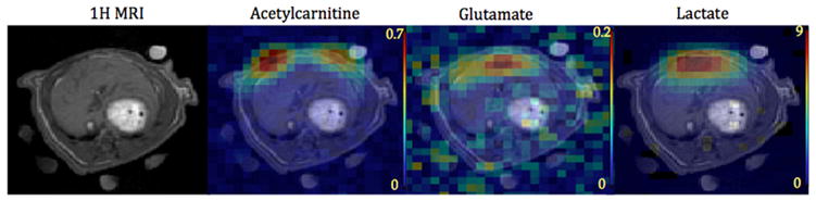
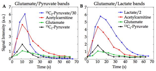
Results: Phantom experiments verified the radiofrequency pulse design and demonstrated that the signal behavior of each group of resonances was unaffected by excitation of the other frequency bands. Dynamic three-dimensional (13) C CSI data demonstrated the sequence capability to image pyruvate, lactate, acetylcarnitine, glutamate, and acetoacetate, enabling the analysis of organ-specific spectra and metabolite time courses.

Conclusions: The presented method allows CSI of widely separated resonances without chemical shift displacement artifact, acquiring multiple frequency bands alternately to obtain dynamic time-course information. This approach enables robust imaging of downstream metabolic products of acetyl-coenzyme A with hyperpolarized [2-(13) C]pyruvate.

Keywords: [2-13C]pyruvate; dynamic metabolic imaging; hyperpolarized 13C; spiral CSI.


Figures

Figure 1
Figure 1
The spectrally selective hamming windowed-sinc RF pulse (Fig. A) and its spectral profile in log scale with passband placed on the glutamate band (Fig. B) and on the pyruvate band (Fig. C). The dashed lines marked G indicate the chemical shifts of [5-13C]glutamate and [1-13C]pyruvate in the passband with the resonances of acetylcarnitine, acetoacetate and citrate located between them. With the passband placed on the glutamate band, signal from [2-13C]pyruvate (line P) is suppressed. Lines L and A mark the locations of [2-13C]lactate and [2-13C]alanine, respectively. To image [2-13C]pyruvate or [2-13C]lactate, the passband is shifted to the corresponding frequency.
Figure 2
Figure 2
(A) 1H SPGR MRI and overlaid 13C metabolic images of a coronal slice (reformatted from 3D axial acquisition) through the syringe containing co-polarized [2-13C]pyruvate and [1-13C]lactate solution. (B) Comparison of in vitro T1 measurements for a co-polarized solution of [2-13C]pyruvate and [1-13C]lactate when both resonances were imaged in an interleaved manner (solid blue line) with the excitation/acquisition of each band individually (dashed red line) using 3D CSI with frequency band-selective excitation. The similar T1 values obtained demonstrate that the excitation of one band did not affect the signal behavior of resonances in the other band.
Figure 3
Figure 3
(left) Spectrum of the glutamate frequency band averaged over all time-points acquired from rat heart in vivo showing the 13C resonances of glutamate (Glu), acetoacetate (Aca), acetylcarnitine (Alcar) as well as natural abundance [1,2-13C]pyruvate doublet (13C1Pyr) in the injected solution. The dashed line near 170 ppm indicates [2-13C]pyruvate signal excited by RF stopband ripples and aliased into the glutamate band. Citrate at 181 ppm was not reliably detected with sufficient SNR and may overlap with signal from [1-13C]pyruvate hydrate. (right) Time-averaged spectrum of the lactate band acquisition showing the [2-13C]lactate doublet acquired from the same rat. The RF passband was centered near the down-field peak of the doublet to avoid signal contamination from [2-13C]alanine resulting in slightly different flip angles for the two peaks.
Figure 4
Figure 4
Time-averaged 13C metabolite maps from a bolus injection of hyperpolarized [2-13C]pyruvate superimposed on to 1H MR images for one rat. The central slice of a 3D acquisition centered on the heart is shown. The images were acquired at 10 min after dichloroacetate infusion, except for [2-13C]lactate, which was acquired 2 h post-DCA from a separate [2-13C]pyruvate injection.
Figure 5
Figure 5
Time-averaged 13C metabolite maps of a slice through the liver superimposed on to 1H MR images. A central slice from a 3D acquisition centered on the liver is shown. The images were averaged from two injections of hyperpolarized [2-13C]pyruvate. Only the glutamate band and lactate band were imaged and the pyruvate band was not imaged in this case. The acetylcarnitine resonance lies within the glutamate frequency band.
Figure 6
Figure 6
Metabolite signal time-courses from heart ROI after bolus injections of hyperpolarized [2-13C]pyruvate with alternate acquisition of (A) the glutamate frequency band and the [2-13C]pyruvate band or (B) the glutamate band and the [2-13C]lactate band. The glutamate, acetylcarnitine and [1-13C]pyruvate signals are from the glutamate band acquisition. The SNR of acetoacetate was insufficient to plot time-courses. The 13C1-pyruvate signal is from the 13C1-13C2 doublet from natural abundance [1,2-13C]pyruvate in the injected [2-13C]pyruvate solution.

References

    1. Kurhanewicz J, Vigneron DB, Brindle K, Chekmenev EY, Comment A, Cunningham CH, Deberardinis RJ, Green GG, Leach MO, Rajan SS, Rizi RR, Ross BD, Warren WS, Malloy CR. Analysis of cancer metabolism by imaging hyperpolarized nuclei: prospects for translation to clinical research. Neoplasia. 2011;13(2):81–97. - PMC - PubMed
    1. Malloy CR, Merritt ME, Dean Sherry A. Could 13C MRI assist clinical decision-making for patients with heart disease? NMR Biomed. 2012;24(8):973–979. - PMC - PubMed
    1. Brindle KM, Bohndiek SE, Gallagher FA, Kettunen MI. Tumor imaging using hyperpolarized 13C magnetic resonance spectroscopy. Magn Reson Med. 2011;66(2):505–519. - PubMed
    1. Golman K, Zandt RI, Lerche M, Pehrson R, Ardenkjaer-Larsen JH. Metabolic imaging by hyperpolarized 13C magnetic resonance imaging for in vivo tumor diagnosis. Cancer Res. 2006;66(22):10855–10860. - PubMed
    1. Kohler SJ, Yen Y, Wolber J, Chen AP, Albers MJ, Bok R, Zhang V, Tropp J, Nelson S, Vigneron DB, Kurhanewicz J, Hurd RE. In vivo 13carbon metabolic imaging at 3T with hyperpolarized 13C-1-pyruvate. Magn Reson Med. 58(1):65–69. 207. - PubMed

Publication types

MeSH terms

LinkOut - more resources