. 2013 Sep;163(1):431-40.
doi: 10.1104/pp.113.221713. Epub 2013 Jul 22.

Histone deacetylase AtHDA7 is required for female gametophyte and embryo development in Arabidopsis

Affiliations

Histone deacetylase AtHDA7 is required for female gametophyte and embryo development in Arabidopsis

Riccardo Aiese Cigliano et al. Plant Physiol. 2013 Sep.

Abstract

Histone modifications are involved in the regulation of many processes in eukaryotic development. In this work, we provide evidence that AtHDA7, a HISTONE DEACETYLASE (HDAC) of the Reduced Potassium Dependency3 (RPD3) superfamily, is crucial for female gametophyte development and embryogenesis in Arabidopsis (Arabidopsis thaliana). Silencing of AtHDA7 causes degeneration of micropylar nuclei at the stage of four-nucleate embryo sac and delay in the progression of embryo development, thereby bringing the seed set down in the Athda7-2 mutant. Furthermore, AtHDA7 down- and up-regulation lead to a delay of growth in postgermination and later developmental stages. The Athda7-2 mutation that induces histone hyperacetylation significantly increases the transcription of other HDACs (AtHDA6 and AtHDA9). Moreover, silencing of AtHDA7 affects the expression of ARABIDOPSIS HOMOLOG OF SEPARASE (AtAESP), previously demonstrated to be involved in female gametophyte and embryo development. However, chromatin immunoprecipitation analysis with acetylated H3 antibody provided evidence that the acetylation levels of H3 at AtAESP and HDACs does not change in the mutant. Further investigations are essential to ascertain the mechanism by which AtHDA7 affects female gametophyte and embryo development.


Figures

Figure 1.
Figure 1.
Schematic representation of the AtHDA7 gene structure and its promoter region. The dark gray triangle indicates the position of the Transfer-DNA in Salk_002912C.
Figure 2.
Figure 2.
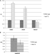
Expression analysis of AtHDA7 and the level of acetylated H3 in Athda7-2 leaf. A, Quantitative RT-PCR. The fold change values are averages of three replicates. The se is reported as a black vertical bar. B, Immunoblotting performed by anti-H3K9K14Ac and anti-H3 antibodies. The relative ratio of H3K9K14Ac/H3 and se (three replicates) are reported in the bar diagram. *P < 0.05. WT, Wild type.
Figure 3.
Figure 3.
Seed germination in Athda7oe (n = 124) and Athda7-2 (n = 316) as compared with the wild type (wt; n = 125). Radicle emergence (stage 0.5 according to Boyes et al., 2001) or later stages were recorded at reported days relative to the date of seed incubation at 24°C. Seeds were preincubated at 4°C for 4 d. Average values are shown with sd. *P ≤ 0.001.
Figure 4.
Figure 4.
In vitro growth stages of seedlings in Athda7oe (n = 114) and Athda7-2 (n = 277) compared with the wild type (n = 120). The frequency of seedlings at stages from 0.5 to 1.04 (according to Boyes et al., 2001) was recorded at reported days relative to the date of seed incubation at 24°C. Seeds were preincubated at 4°C for 4 d. The values are averages of three replicates. *P ≤ 0.05.
Figure 5.
Figure 5.
In soil growth of Athda7oe (n = 37) and Athda7-2 (n = 40) compared with the wild type (wt; n = 35). The number of rosette leaves was recorded over a period of 32 d after sowing (DAS). Flowering time is indicated by arrows. *P ≤ 0.05, **P ≤ 0.01, ***P ≤ 0.001.
Figure 6.
Figure 6.
Silique fertility in Athda7oe and Athda7-2 compared with the wild type (wt). Average values estimated on 20 siliques are shown with sd.
Figure 7.
Figure 7.
Ovule and female gametophyte development in the wild type (A–F) and Athda7-2 (G–L). A and G, One-nucleate female gametophyte at stage FG1 (according to Christensen et al., 1997), indicated by the arrows. Degenerate megaspores are present. B and H, Two-nucleate female gametophyte at stage FG2, indicated by the arrows. C, D, and I, Four-nucleate female gametophyte at late stage FG4. Two pairs of nuclei at each pole are separated by a large central vacuole, and the chalazal nuclei are positioned along a line that is parallel to the chalazal-micropylar axis. J, Female gametophyte at late stage FG4 showing micropylar nuclei in degeneration, indicated by the arrow. E and K, Eight-nucleate/seven-celled mature female gametophyte. Observations were performed by DIC (C, E, I, and K) and phase-contrast microscopy. The female gametophyte in A, B, D, E, and G is oriented with its micropylar pole to the left, while the micropylar pole in C, H, I, J, and K is at right. F and L, Ovules excised from prefertilization pistils and observed by fluorescence microscopy through DAPI staining. In Athda7-2, the ovule is degenerating. Bars = 20 µm. [See online article for color version of this figure.]
Figure 8.
Figure 8.
Embryogenesis in the wild type (A) and Athda7-2 (B). A, All embryos are at the heart stage. B, One embryo is at the heart stage, while the remaining ovules are degenerating. Bars = 200 µm. [See online article for color version of this figure.]
Figure 9.
Figure 9.
Embryo stages in the wild type (Wt) and Athda7-2 at 10 d after pollination. *P < 0.05.
Figure 10.
Figure 10.
Expression analysis of AtHDA6, AtHDA9, and AtHDA19 in Athda7-2 leaf (A) and of AtAESP in flower bud (B) in comparison with the wild type. The fold change (FC) values are averages of three replicates. The se is reported as a black vertical bar. *P < 0.05.

References

    1. Alexander MP. (1969) Differential staining of aborted and nonaborted pollen. Stain Technol 44: 117–122 - PubMed
    1. Alonso JM, Stepanova AN, Leisse TJ, Kim CJ, Chen H, Shinn P, Stevenson DK, Zimmerman J, Barajas P, Cheuk R, et al. (2003) Genome-wide insertional mutagenesis of Arabidopsis thaliana. Science 301: 653–657 - PubMed
    1. Aufsatz W, Mette MF, van der Winden J, Matzke M, Matzke AJM. (2002) HDA6, a putative histone deacetylase needed to enhance DNA methylation induced by double-stranded RNA. EMBO J 21: 6832–6841 - PMC - PubMed
    1. Barra L, Aiese-Cigliano R, Cremona G, De Luca P, Zoppoli P, Bressan RA, Consiglio MF, Conicella C. (2012) Transcription profiling of laser microdissected microsporocytes in an Arabidopsis mutant (atmcc1) with enhanced histone acetylation. J Plant Biol 55: 281–289
    1. Bell O, Tiwari VK, Thomä NH, Schübeler D. (2011) Determinants and dynamics of genome accessibility. Nat Rev Genet 12: 554–564 - PubMed

Publication types

MeSH terms